Follow-Up Ultrasound after Pyeloplasty should be Performed with An Empty Bladder: A Lesson from Monsieur Laplace
Article Information
Sergio Ghirardo1*, Mario Diplomatico2 , Matteo Zancanaro3, Luca Basso4, Marco Pennesi5 , Egidio Barbi11,5, Federica Pederiva5
1University of Trieste, Trieste, Italy
2Department of Woman, Child and of General and Specialized Surgery, Università degli Studi della Campania "Luigi Vanvitelli", Naples, Italy
3SISSA mathLab, Trieste, Italy
4University of Genova, Genova, Italy
5Institute for Maternal and Child Health - IRCCS “Burlo Garofolo” – Trieste Italy
*Corresponding author: Sergio Ghirardo, University of Trieste, via dell?Istria 65/1, Trieste, Italy, 34137
Received: 27 September 2019; Accepted: 11 October 2019; Published: 18 October 2019
Citation: Sergio Ghirardo, Mario Diplomatico, Matteo Zancanaro, Luca Basso, Marco Pennesi, Egidio Barbi, Federica Pederiva. Follow-Up Ultrasound after Pyeloplasty should be Performed with An Empty Bladder: A Lesson from Monsieur Laplace. Archives of Clinical and Biomedical Research 3 (2019): 357-360.
View / Download Pdf Share at FacebookAbstract
Reason to report: Hydronephrosis caused by congenital ureteropelvic obstruction is quite common affecting up to 0.2% of infants and a pyeloplasty is the way to correct. Ultrasound is the technique of choice to follow up this kind of patient, but no other recommendations are present in literature.
What was unique: So far no one has studied how the changing in the urinary way reflect the ultrasound image. We applied Laplace law to the geometric modelling of the urinary way of ten patients that underwent pyeloplasty before the age of three.
Ramification of this report: After pyeloplasty, due to its reduced thickness, the pelvis presents a tendency to dilate, even by a low increase of urinary pressure.
Ultrasound evaluation with an empty bladder can distinguish between an obstructive pattern and a reoccurrence of junctional occlusion.
Keywords
Hydronephrosis; Pyeloplasty; Follow-Up; Ultrasound; Ureteropelvic Junction Obstruction; Renal Pelvis Dilation; Paediatric; Laplace Law; Mathematical Modelling
Hydronephrosis articles, Pyeloplasty articles, Follow-Up articles, Ultrasound articles, Ureteropelvic Junction Obstruction articles, Renal Pelvis Dilation articles, Paediatric articles, Laplace Law articles, Mathematical Modelling articles
Article Details
1. Introduction
Hydronephrosis caused by congenital ureteropelvic obstruction is quite common affecting up to 0.2% of infants. It is diagnosed thanks to hydronephrosis progression over time at the ultrasound and obstructive pattern at the scintigraphy [1]. To correct obstruction Anderson-Hynes pyeloplasty incises just distally to the pelvic-ureteric junction and the dilated renal pelvis is tapered excising the redundant portion, obtaining a normalisation of the pelvis’ diameter [2]. Such procedure is usually performed on a massively dilated pelvis, and it has no impact on the wall’s thickness which is already markedly reduced. Since the scope of surgery is to preserve the Residual Renal Function, thus the subsequent follow-up is crucial [3]. Considering that the diameter of the pelvis varies considerably with the patient position and bladder status [4], we retrospectively collected urinary-Magnetic Resonance Imaging (urinary-MRI) of ten patients that underwent pyeloplasty before the age of three.
2. Discussion
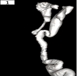
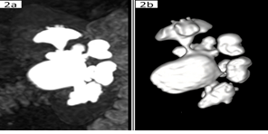
With MRI 3D reconstruction, we found that urethra and ureters could be roughly modelled as a cylinder (Figure 1) while dilated pelvis could be modelled as a sphere (Figure 2a and b).
Consequently, after geometric modeling, we applied Laplace law to both kinds of objects. For the sphere: Wall Tension = (Transmural Pressure × Radius)/(2 × Wall Thickness) and to cylinder: Wall Tension = (Transmural Pressure × Radius)/(Wall Thickness). Wall tension is the force that balances the inner urinary pressure contrasting the dilation and, it is half for a sphere than for a cylinder. Wall tension increases with the radius and it decrease with the thinning of the bladder wall [5]. For this reason, a pelvis that underwent pyeloplasty, due to its reduced thickness, presents a structural tendency to dilate, even by a low increase of urinary pressure. Even, a little increase in pressure may lead to a significative dilation of this “highly compliant” thin pelvis. In this case, pelvis dilation is not associated with renal compression and kidney function is preserved. When the bladder is full, pressure increases in the entirely urinary tracts (pelvis included) and may lead to relevant dilation of pelvis previously operated for hydronephrosis.
3. Conclusion
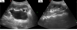
In the follow-up of these patients, discriminating between a nearly physiological dilation caused by a full bladder without an obstructive pattern and a reoccurrence of junctional occlusion that may need a new surgical procedure is crucial. These data suggest that ultrasound evaluation with an empty bladder can distinguish between these two conditions. Indeed, if an obstructive relapse is present, dilation will be persistent regardless of the content of the bladder, while a non-pathologic and non-obstructive dilation will resolve after emptying as shown in Figure 3 (a and b), with no evidence of obstruction at scintigraphy.
Declaration
Ethics approval and consent to participate
No ethical approval was necessary.
Consent for publication
Every author gave the consent for publication.
Availability of data and materials
All clinical documentations can be sent to editors by authors if needed.
Competing interests
There is no competing of interest real or perceived.
Funding
No found was necessary.
Authors' contributions
Sergio Ghirardo, Federica Pederiva and Mario Diplomatico wrote the first draft of the manuscript and they collected data. Matteo Zancanaro performed the geometrical and physical analysis of data. Marco Pennesi and Egidio Barbi designed the study and they made the revision of the manuscript. All authors approved the final version of the manuscript.
Acknowledgements
No acknowledgements are necessary.
References
- Sussman RD, Blum ES, Sprague BM, Majd M, Rushton HG, et al. Prediction of Clinical Outcomes in Prenatal Hydronephrosis: Importance of Gravity Assisted Drainage. J. Urol 197 (2017): 838-844.
- Newling DW, Heslop RW, Kille JN. Pelvioureteral obstruction: results of the Anderson-Hynes pyeloplasty procedure. J. Urol 111 (1974): 12-8.
- Thorup J, Mortensen T, Diemer H, Johnsen A, Nielsen OH. The prognosis of surgically treated congenital hydronephrosis after diagnosis in utero. J. Urol 134 (1985): 914-7.
- Fernbach SK, Bernfield JB. Positional variation in the ultrasound appearance of the renal pelvis. Pediatr. Radiol 21 (1990): 45-7.
- Champmartin S, Ambari A, Le Pommelec JY. New procedure to measure simultaneously the surface tension and contact angle. Rev. Sci. Instrum 87 (2016): 055105.


 Impact Factor: * 3.1
 CiteScore: 2.9
  Acceptance Rate: 11.01%
  Time to first decision: 10.4 days
  Time from article received to acceptance: 2-3 weeks