A Splendid Pelvic Tumor, Indeed
Zilberman DE1, Waldman D2*, Raviv-Zilka L3*, Fridman E4, Ben-Shlush A3*, Mor Y1*
1Departments of Urology, The Chaim Sheba Medical Center and Edmond and Lily Safra Children's Hospital, Tel-Hashomer, Ramat-Gan, Affiliated to Sackler School of Medicine, Tel-Aviv University, Israel
2Pathology, The Chaim Sheba Medical Center and Edmond and Lily Safra Children's Hospital, Tel-Hashomer, Ramat-Gan, Affiliated to Sackler School of Medicine, Tel-Aviv University, Israel
3Pediatric Hemato-Oncology, The Chaim Sheba Medical Center and Edmond and Lily Safra Children's Hospital, Tel-Hashomer, Ramat-Gan, Affiliated to Sackler School of Medicine, Tel-Aviv University, Israel
4Unit of Pediatric Imaging, The Chaim Sheba Medical Center and Edmond and Lily Safra Children's Hospital, Tel-Hashomer, Ramat-Gan, Affiliated to Sackler School of Medicine, Tel-Aviv University, Israel
*Corresponding Author: Prof. Yoram Mor, Departments of Urology, The Chaim Sheba Medical Center and Edmond and Lily Safra Children's Hospital, Tel-Hashomer, Ramat-Gan, Affiliated to Sackler School of Medicine, Tel-Aviv University, Israel
Received: 27 May 2020; Accepted: 18 June 2020; Published: 02 July 2020
Article Information
Citation: Zilberman DE, Waldman D, Raviv-Zilka L, Fridman E, Ben-Shlush A, Mor Y. A Splendid Pelvic Tumor, Indeed. Archives of Nephrology and Urology 3 (2020): 046-048.
DOI: 10.26502/anu.2644-2833020
View / Download Pdf Share at FacebookAbstract
Accessory spleen is not a common finding, usually located nearby the normal anatomic location of the spleen, oftentimes in the splenic hilum, the great omentum and the pancreas. Pelvic accessory spleen is a very rare finding, mostly asymptomatic and incidentally radiologically detected. Herein, we present an 18 years old male who underwent an investigation for daytime urinary frequency and suspicious small right pelvic mass was demonstrated by both ultrasound and MRI scans. In view of being a potentially malignant tumor, a robotic-assisted removal was uneventfully performed and the final pathology was surprisingly compatible with an accessory spleen.
Keywords
<p>Accessory spleen; Pelvis; Pelvic accessory spleen</p>
Article Details
1. Case Report
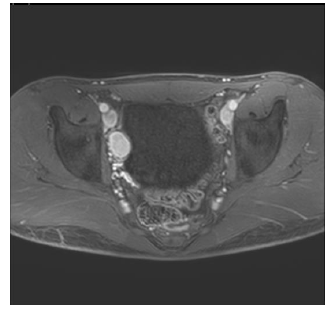
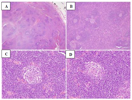
An 18 years old generally healthy male presented with daytime urinary frequency. Physical examination and urinalysis were unremarkable. Abdominal ultrasound (US) demonstrated a solid lesion, 3 cm in diameter, adjacent to the Rt. Bladder wall. Magnetic resonance imaging (MRI) (Figure 1) demonstrated a similar finding, namely, a solid homogenous enhancing mass on T1 weighted images after injection of Gadolinium (26 × 18 mm), pressuring the upper-lateral aspect of the urinary bladder. In view of a potentially malignant tumor, a robotic-assisted removal of the tumor was uneventfully performed. The final pathology was compatible with an accessory spleen showing reactive lymphoid follicular hyperplasia (Figure 2). A repeat US, obtained 6 months postoperatively showed disappearance of the finding, and the patient remained well at 6-months follow-up visit.

Figure 1: Abdominal MRI showing a solid homogenous enhancing mass (26 × 18 mm), on T1 weighted images after injection of Gadolinium, pressuring the upper-lateral aspect of the urinary bladder.

Figures 2: A - H&E x 2 Normal splenic tissue surrounded by splenic capsule; B - H&E x 4 – Splenic tissue with reactive lymphoid follicles; C, D - H&E x 40 - Central arteriole in a reactive lymphoid follicle.
2. Discussion
Ectopic splenic tissue can be encountered either due to auto-transplantation of cells following abdominal trauma or splenectomy (splenosis), or as a congenital malformation (accessory spleen) [1]. Accessory spleens were reported in 6.7% of autopsies, mainly in the splenic hilum, the great omentum and the pancreas [2]. However, pelvic accessory spleen is a very rare finding, mostly asymptomatic and incidentally radiologically detected, though it may cause acute abdominal pain, abdominal discomfort, or dysmenorrhea [3-4]. It can be well visualized by either Computerized Tomography (CT) scans, MRI which often shows a vascular pedicle originating from the great omentum or from the splenic hilum, as well as scintigraphy with Tc-99m-labeled colloids which localize in the reticuloendothelial cells of the liver and spleen [5]. Yet, as it mimics a primary tumor or a metastatic lymph node, it is usually removed to avoid concern.
Conflicts of Interest
The authors declare that they have no competing interests.
Financial Disclosure
The authors have no proprietary or commercial interests.
References
- Taskin MI, Baser BG, Adali E, et al. Accessory spleen in the pelvis: A case report. Int. J. Surg Case Rep 12 (2015): 23-25.
- Tendler R., Farah RK, Kais M, et al. Symptomatic pelvic accessory spleen in a female adolescent: Case report. J. Clin. Ultrasound 45 (2007): 600-602.
- Ota H, Ojima Y, Sumitani D, et al. Dynamic computed tomography findings of an accessory spleen in the pelvis: a case report. Surg. Case Rep 2 (2016): 2016.
- Zhou JS, Chen X, Zhu T, et al. Pelvic accessory spleen caused dysmenorrhea. Taiwan J. Obstet. Gynecol 54 (2015): 445-446.
- Iorio F, Frantellizzi V, Drudi FM, et al. Locally vascularized pelvic accessory spleen. J. Ultrasound 19 (2016): 141-144.


 Impact Factor: * 3.3
 Acceptance Rate: 73.59%
  Time to first decision: 10.4 days
  Time from article received to acceptance: 2-3 weeks