Assessment of Smile in Post-Orthodontic Patients using Smilecurve Templates- A Retrospective study
Chhavi G*, Divya S, Bhupender S, Payal S
Department of Orthodontics and Dentofacial Orthopedics at I.T.S Dental College, Muradnagar, Ghaziabad, India
*Corresponding Author: Chhavi Grover, Department of Orthodontics and Dentofacial Orthopedics at I.T.S Dental College, Muradnagar, Ghaziabad, India
Received: 30 August 2023; Accepted: 15 September 2023; Published: 21 September 2023
Article Information
Citation: Chhavi Grover, Divya Shetty, Bhupender Singh, Payal Sharma. Assessment of Smile in Post- Orthodontic Patients using Smilecurve Templates- A Retrospective study. Dental Research and Oral Health. 6 (2023): 58-64.
View / Download Pdf Share at FacebookAbstract
Aim: The aim of this article was to assess changes in smile aesthetics using SmileCurve Templates in patients treated with fixed mechanotherapy.
Materials and Methods: Pre-treatment and Post Debonding photographs of 30 patients were taken and SmileCurve Template (SCT) was superimposed on the Front Occlusal photographs of the patient to check for correction in the Smile aesthetics from Pre-treatment to Post-treatment. The six criteria in the Diagram of Dental Aesthetic Reference (DDAR) that included Symmetry, Tooth axes, Gingival contour, Interproximal contacts, Incisal edges, Dental proportions were evaluated. Among the parameters of horizontal lines only two criteria that is the Cervical line and Papillary line were evaluated. The pre and post treatment frontal occlusal photographs were evaluated for the above mentioned 6 DDAR criteria and 2 horizontal lines.
Results: The post-treatment smile showed a statistically significant improvement as compared to the pre- treatment smile.
Conclusion: There was improvement in the various parameters influencing smile aesthetics. The SmileCurve template is a useful tool that can help clinicians to objectively assess the parameters influencing smile aesthetics and take corrective measures, as required.
Keywords
<p>Smile, SmileCurve Templates, DDAR</p>
Article Details
Introduction
Facial appearance more specifically smile aesthetics, plays a preponderant role. Smile is considered to be an essential facial expression that plays an important role in social interactions. The aim of orthodontic treatment is not merely the correction of dentition but to achieve a favourable esthetics of face. Esthetically pleasing smile enhances attractiveness thus contributes in boosting one’s confidence [1]. Thus, inspired by pretty faces and beautiful smiles, patients have sought treatment modalities to improve dentofacial esthetics and yield positive changes in their smile [2]. There are several reference parameters that are to be followed to achieve an esthetic smile. Golden proportion, golden percentage and recurring esthetic dental are theories introduced in this field. Lombardi was the first to suggest the application of the golden proportion in dentistry and Levin suggested the use of the theory of Golden proportion to relate the successive width of the anterior teeth, as viewed from the labial aspect. Ward suggested the recurring esthetic dental (RED) proportion. Snow considered a bilateral analysis of apparent individual tooth width as a percentage of the total apparent width of the six anterior teeth. He proposed the golden percentage, wherein the proportional width of each tooth should be: canine 10%, lateral 15%, central 25%, central 25%, lateral 15%, and canine 10% of the total distance across the anterior segment, in order to achieve an esthetically pleasing smile [3]. The use of simple and reliable mechanisms can improve the possibilities of success, if not eliminate performance errors. The Diagram of Facial Aesthetic References (DFAR) is an auxiliary diagnostic tool that is well suited to that purpose. The diagram consists of six frames that surround the maxillary incisors and canines; their limits are specific to each aesthetic reference. The function of the DFAR is to give an exact idea of the positioning and ratios between teeth, as well as their relationship with the gum and lips [4]. For that purpose, Camara [4] in 2020 designed the SmileCurves template which was designed by using the Diagrams of Dental Aesthetic References (DDAR), together with the Six Horizontal Smile Lines. These two analytical frameworks alone include a number of variables, enough to build a guide for the correct positioning of anterior maxillary teeth in a beautiful smile [5]. The aim of this article was to assess changes in smile aesthetics using SmileCurve Templates in patients treated with fixed mechanotherapy.
Materials and Methods
Materials:
- Pre Frontal occlusal photographs
- Post Debonding Frontal occlusal photographs
- DSLR Camera
- Picasa Photo editor
- Cheek Retractors
- Laptop
Inclusion criteria:
- Age group of 12-25 years
- Permanent dentition
- Fully erupted anterior teeth
- No smoking habit
- No history of any drug intake
- Patients who underwent Fixed Orthodontic therapy with same MBT Bracket system and 0.022 slot dimension.
Exclusion criteria:
- Patients with systemic problems
- Periodontal problems
Criteria for standardization of photographs
- All photographs were taken with a natural head posture.
- Only frontal occlusal photographs were evaluated.
- The photographs were taken using Nikon D5600 DSLR camera.
- All the photographs were cropped using Picasa 3 to a standard size and 4x6 ratio.
Method
Pre-treatment and Post Debonding photographs of 30 patients were taken and SmileCurve Template (SCT) was superimposed on the Front Occlusal photographs of the patient to check for correction in the Smile aesthetics from Pre-treatment to Post-treatment.
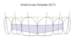
Figure 1: Smile Curve Template
The six criteria in the Diagram of Dental Aesthetic Reference (DDAR) were Symmetry, Tooth axes, Gingival contour, Interproximal contacts, Incisal edges, Dental proportions. Among the parameters of horizontal lines only two criteria that is the Cervical line and Papillary line were evaluated. The pre and post treatment frontal occlusal photographs were evaluated for the above mentioned 6 DDAR criteria and 2 Horizontal lines.
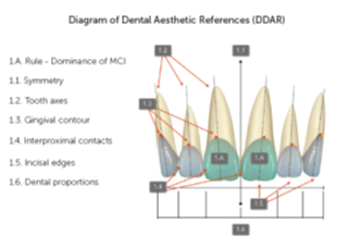
Figure 2: Diagram of Dental Aesthetic Reference (DDAR)
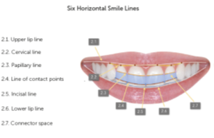
Figure 3: Six Horizontal Smile Lines
SCT was used by adapting the template in a PNG file (semi-transparent) over images of JPEG, TIFF or STL files, or any other image files. The rule of dominance was applied to the photographs and the width of maxillary central incisors coincided on frontal smile or intraoral image, and the coincidences, or lack of them, was evaluated using the DDAR references and the six horizontal smile lines. The proportions of the photographs were kept constant i.e. 4x6. A nominal scale (Yes/No) of measurement was used. Only the 6 anterior teeth were evaluated. For a particular criterion, Yes was designated if 3 or more than 3 out of 6 teeth coincided whereas No was designated for any value that was less than 3. The distribution of patients according to the extent of concordance with the SCT parameters was evaluated.
After evaluation
- If all the parameters were coinciding in Smile photographs with the SCT then 100% concordance was seen.
- If one parameter was not coinciding in Smile photographs with the SCT then 87.5% concordance was seen.
- If 2 parameters were not coinciding in Smile photographs with the SCT then 75% concordance was seen.
- If 3 parameters were not coinciding in Smile photographs with the SCT then 62.5% concordance was seen.
- If 4 parameters were not coinciding in Smile photographs with the SCT then 50% concordance was seen.
- If 5 or more parameters were not coinciding in Smile photographs with the SCT then 37.5% concordance was seen.
The concepts of DDAR and the six horizontal smile lines is the bases of the SmileCurves template, a tool to facilitate the evaluation of dental and oral aesthetics. SCT is composed of diagrams that represent the six maxillary anterior teeth, and the principal elements are the central incisors. The drawing of these teeth aims to help in the evaluation and visualization of dental and gingival contours.
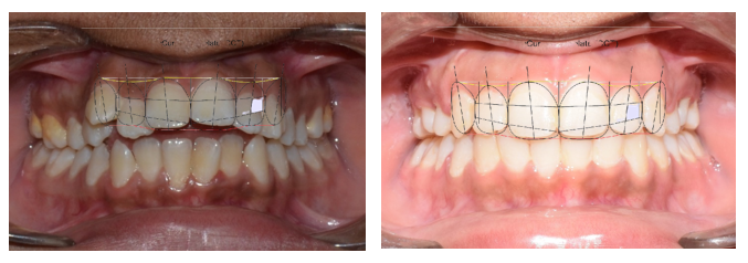
Figure 4: Pre and Post Treatment Photographs
Results
Table 1 and graph 1 show Distribution of patients (number and percentage) according to concordance with SCT parameters. The comparisons showed that the data was significant and table 1 showed that five or more criteria were not met in twenty four patients before treatment and in two patients after the treatment. Four criteria were not met in two patients before treatment and three samples after the treatment. Three criteria were not met in one sample before the treatment and one sample after the treatment. Two criteria were not met in one sample before the treatment and three samples after the treatment. Four criteria were not met in one sample after the treatment. All the criteria were met in two samples before the treatment and twenty samples after the treatment.
|
Concordance Time |
100% |
87.5% |
75% |
62.5% |
50% |
37.5% or less |
Total |
|
Pre-Treatment Smile Photographs |
2 (6.7%) |
0 (0.0%) |
1 (3.3%) |
1 (3.3%) |
2 (6.7%) |
24 (80%) |
30 |
|
Post-Treatment Smile Photographs |
20 (66.7%) |
1 (3.3%) |
3 (10%) |
1 (3.3%) |
3 (10%) |
2 (6.7%) |
30 |
Table 1: Distribution of Patients (number and percentage) according to concordance with SCT parameters
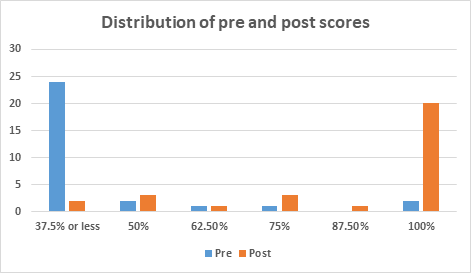
Graph 1: Distribution of Patients (percentage) according to concordance with SCT parameters
|
Interval |
Mean |
SD |
Difference |
p value |
95% CI of difference |
|
|
Lower |
Upper |
|||||
|
Pre |
44.58 |
17.27 |
42.09 |
0.001* |
-52.08 |
-32.09 |
|
Post |
86.67 |
21.51 |
||||
Table 2: Comparison of pre and post treatment distribution of patients
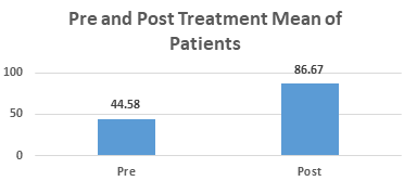
Graph 2: Comparison of pre and post treatment distribution of patients
Table 2 and graph 2 showed the Comparison of pre and post treatment distribution of patients. The pre-treatment mean ranged from 44.58 ± 17.27 while post-treatment mean ranged from 86.67 ± 21.51. The p-value of 0.001 suggested a significant difference in the pre-treatment and post-treatment mean.
|
Parameter |
Pre |
Post |
p value |
|
|
Symmetry |
Yes |
12 (40%) |
17 (56.7%) |
0.458 (NS) |
|
No |
18 (60%) |
13 (43.3%) |
||
|
Tooth axis |
Yes |
13 (43.3%) |
16 (53.3%) |
0.710 (NS) |
|
No |
17 (56.7%) |
14 (46.7%) |
||
|
Incisal edge |
Yes |
5 (16.7%) |
21 (70%) |
0.003* |
|
No |
25 (83.3%) |
9 (30%) |
||
|
Tooth position |
Yes |
11 (36.7%) |
18 (60%) |
0.265 (NS) |
|
No |
19 (63.3%) |
12 (40%) |
||
|
Gingival contour |
Yes |
2 (6.7%) |
19 (63.3%) |
0.001* |
|
No |
29 (93.3%) |
11 (36.7%) |
||
|
Interproximal contacts |
Yes |
2 (6.7%) |
23 (76.7%) |
0.001* |
|
No |
29 (93.3%) |
7 (23.3%) |
||
|
Cervical line |
Yes |
5 (16.7%) |
17 (56.7%) |
0.017* |
|
No |
25 (83.3%) |
13 (43.3%) |
||
|
Papillary line |
Yes |
4 (13.3%) |
21 (70%) |
0.001* |
|
No |
26 (86.7%) |
9 (30%) |
McNemar test; * indicates significant difference at p≤0.05
Table 3: Comparison of distribution of patients from pre to post treatment in each parameter
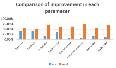
Graph 3: Comparison of distribution of patients from pre to post treatment in each parameter
Table 3 and graph 3 shows significant improvement post treatment was seen in following parameters: incisal edge, gingival contour, interproximal contacts, cervical line and papillary line.
|
Parameter |
Improvement (in %) |
|
Symmetry |
16.70% |
|
Tooth Axis |
10.00% |
|
Incisal Edge |
53.30% |
|
Tooth Position |
23.30% |
|
Gingival Contour |
56.60% |
|
Interproximal Contacts |
70.00% |
|
Cervical Line |
40.00% |
|
Papillary Line |
56.70% |
Table 4: Evaluation of improvement (in %) for pre to post treatment in each parameter among study subjects
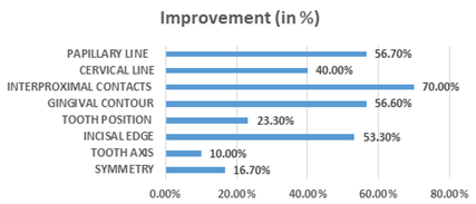
Graph 4: Evaluation of improvement (in %) for pre to post treatment in each parameter among study subjects
Table 4 and graph 4 shows maximum improvement was seen in interproximal contacts (70%) followed by papillary line (56.70%), gingival contour (56.60%) and incisal edge (53.30%). Least improvement was seen in symmetry (16.70%) and tooth axis (10.00%).
Discussion
The present study aassessed the Smile in Post-Orthodontic patients using SmileCurve Templates in Indian population. The sample in the study consisted of thirty patients who underwent fixed orthodontic therapy. Frontal occlusal photographs of the patients were taken and SmileCurve templates was superimposed on the same. The template placement was based on the rule of dominance i.e the Central incisors. The six criteria in the DDAR reference that were Symmetry, Tooth axes, Gingival contour, Interproximal contacts, Incisal edges, Dental proportions and for horizontal lines only two criteria the Cervical line and Papillary line were evaluated. The concepts of DDAR and the six horizontal smile lines are the bases of the SmileCurves template, a tool to facilitate the evaluation of dental and oral aesthetics. SCT is composed of diagrams that represent the six maxillary anterior teeth, and the principal elements are the central incisors. In other words, DDAR does not use standard measurements, but rather, the subjective perception of agreeability of proportions, supported by the objectivity of the size of boxes [5]. In this study, a nominal scale (Yes/No) of measurement was used. Only the 6 anterior teeth were evaluated. For a particular criterion, Yes was designated if 3 or more than 3 out of 6 teeth coincided whereas No was designated for any value that was less than 3. The pre-treatment and post-treatment mean showed a statistically significant difference. The results of the study showed that pre-treatment most of the parameters evaluated did not coincide with the SmileCurve Template in the smile photographs and 5 or more criteria did not coincide in 24 (80%) patients out of 30 patients. Four criteria were not met in 2 (6.7%) patients whereas 3 and 2 criteria did not coincide in 1 patient (3.3%). There were no patients who had 1 criterion coinciding. Only 2 (6.7%) out of 30 patients had all the parameters that were coinciding. The post-treatment smile showed a statistically significant improvement as compared to the pre- treatment smile. As compared to the pre-treatment values, only 2 (6.7%) patients were there who had 5 or more criteria that were not coinciding. Three patients (10%) each out of 30 were there in the category of 4 and 2 criteria coinciding respectively. Only 1 patient (3.3%) each had 3 and 1 criteria coinciding respectively. Among 30 patients, 20 patients post-treatment had all the parameters that were coinciding with the SmileCurve Template. Considering the criterion of Dental Proportion, Singh et al. in their review article stated that for an esthetically pleasing smile according to the golden proportion, the apparent width of the lateral incisor should be 62% of the width of the central incisor, the apparent width of the canine should be 62% that of the lateral incisor and the apparent width of the first premolar should be 62% that of the canine. This ratio serves as a guide to determine the post-treatment size of a lateral size in case of disproportionately small lateral incisors, or when canines are reduced to replace the lateral incisors in case of congenitally missing laterals [6]. According to Camara, the ideal proportion between height and width of MCI is about 80%. That is, when width is 8.0 mm, height is 10.0 mm. Based on this, dimensions follow the diagonal proportion of a square (1:1.414), which means that the width of lateral incisors corresponds to 71% of the width of central incisors; and that of the canines, 71% of that of the lateral incisors (regressive proportion). This proportion was chosen because dentists, academics and laypeople prefer it.5 In our study the dental proportion improved by 23.30%.
In a study done by Brandão and Brandão, concluded that for tooth axes two proportions must be considered: the relation between height and width of each tooth, and the relation of height and width among the teeth [7]. Gillen et al. investigated dental proportions and found following proportions of width among the upper anterior teeth: a) lateral incisors have 78% of the width of the central incisor (lateral incisor= central incisor x 0.78); b) lateral incisor has 87% of the width of the canine ; c) canine has 90% of the width of the central incisor [8]. The studies by Sterrett et al. demonstrated that the relation between width and height of central and lateral incisors and canines is practically the same in both genders. Females tend to have slightly wider teeth in comparison to males, being: central incisors 86% and 85%, lateral incisors 79% and 76%, and canines 81% and 76%, respectively. The most recent research available found the proportion between height and width of upper anterior teeth ranging from 75 to 80% in central incisors, from 66 to 70% in lateral incisors, and from 80 to 85% in canines, without statistical difference between men and women. Most authors define the height/width ratio of 0.80 for the upper central incisor as a standard [7]. In our study the tooth axes improved by 10%. Pinho et al. in their study on impact of dental asymmetries on the perception of smile esthetics evaluated the horizontal continuity of the gingival contour, teeth, and upper lip and considered them essential for esthetics. They concluded that asymmetries of up to 1.5 mm in the free gingival margin of the maxillary central incisors seemed to be acceptable and not perceived by the laypersons [9]. In our study the tooth symmetry improved by 16.07%. Camara gave the DDAR in which he described incisal edges, interproximal contacts, cervical line, papillary line. The incisal line follows the edges of anterior maxillary teeth. The ideal is that in young patients the incisal edges of the central incisors be below the edges of the lateral incisors and canines in a frontal view. In that configuration, the form of the incisal line resembles the outline of a “deep plate” [4]. In relation to the incisal line, SCT respects the perspective and consequent parallax effect, thus creating a 0.8-mm step between the incisal edges of central and lateral incisors and positions the incisal and occlusal edges of central incisors and canines on the same perspective, creating an incisal line in the shape of a "soup plate" [5]. Incisal edges improved by 53.30% in this study. The interproximal contacts in the maxillary anterior teeth were evaluated starting from Canine in a descending manner. The contact between the canine and lateral incisor is positioned higher than the contact between the lateral and central incisors; the contact between the central incisors is even lower. The contact points should be narrow, unless there is a discrepancy in the mesio-distal diameter of the crown. The position of the contact between teeth is related to tooth position and form. As such, the line that unites these points will be parallel to the incisal line, whenever there is no discrepancy between the sizes, shapes and angles of anterior teeth [5]. In a study done by M.R. Rayyan results showed that the ‘ideal’ interproximal contact area ratio arrangement proposed by Morley and Eubank – 50:40:30 – was considered the most attractive among dentists and dental technicians. Also, in examining the ratio of the length of the interproximal contact areas, the majority of the participants ranked the ‘ideal’ ratio (50:40:30) as the most attractive. One exception was the rating of the male smile by the layperson group, where a higher percentage found the ‘reduced’ ratio (40:30:20) to be the most attractive [10]. The interproximal contacts improved by 70%. The papillary line was formed by the tips of the gingival papillae located between the canines and lateral incisors, and between the maxillary lateral incisors and central incisors. According to Camara, there were no studies that evaluated the standard height for this relationship but based on works that evaluated the ideal height of the central incisors and the relationship between the papillary tips and the position and size of teeth, it was presumed that an ideal line would be parallel to the line formed by the contact points [5]. In this study the papillary line improved by 56.70%. According to the work of Kurt and Kokich [11], the papilla in the central incisors fills half the size of those teeth, under normal conditions and it was expected that the same pattern would be repeated for the lateral incisors and canines. Camara also described an ideal cervical line by the union of the apexes of the canines, maxillary lateral and central incisors. As the most apical point of the gingival contour, the apex in maxillary teeth is usually located distal to the long axis of the tooth. He stated that the apexes of the maxillary canines are most often higher than the lateral incisors and about the same level as the central incisors, the cervical line attains a convex aspect in relation to the occlusal plane which is ideal form of the cervical line. The gingival zeniths of the central incisors and canines have the same height, whereas those of the lateral incisors are a little below (0.6 mm) them [5]. Singh et al. stated that the central incisor and canine gingival margin are at the same level, while the lateral incisor margin is 1.5 mm lower [6]. The cervical line and the gingival contour improved by 40% and 56.60% respectively. A significant improvement post treatment was seen in incisal edge, gingival contour, interproximal contacts, cervical line and papillary line and maximum improvement was seen in interproximal contacts (70%) followed by papillary line (56.70%), gingival contour (56.60%) and incisal edge (53.30%). Least improvement was seen in symmetry (16.70%) and tooth axis (10.00%). This topic of study is unique as there is less evidence on the same topic. This result changed greatly from pre-treatment to post-treatment indicating the usefulness of the SCT in planning of treatment mechanics. Moreover, these templates also provide an effortless and economical tool for the aesthetic analysis of a smile that can be used by a practitioner for smile evaluation. More research is needed to confirm if such an association among different characteristics of smile can be established. One limitation of the present study lies in its lack of generalizability of the results to the whole population.
Conclusions
- There was improvement in the various parameters influencing smile aesthetics like the incisal edge, gingival contour, interproximal contacts, cervical line and papillary line.
- Maximum improvement was seen in interproximal contacts (70%) followed by papillary line (56.70%), gingival contour (56.60%) and incisal edge (53.30%).
- Least improvement was seen in symmetry (16.70%) and tooth axis (10.00%).
- There was substantial improvement in the smile aesthetics with Orthodontic Therapy.
- The SmileCurve template is a useful tool that can help clinicians to objectively assess the parameters influencing smile aesthetics and take corrective measures, as required.
References
- Singh S, Jasoria G, Kushwah A, et al. Macroesthetic Elements of Smile. A Review Article. TMU J Dent 5 (2018): 23-26.
- Machado AW. Ten commandments of smile esthetics. Dental Press J Orthod 19 (2014): 136-157.
- Murthy BVS, Ramani N. Evaluation of natural smile: Golden proportion, RED or Golden percentage. J Conserv Dent 11 (2008): 16-21.
- Câmara CA. Esthetics in Orthodontics: six horizontal smile lines. Dental Press J. Orthod 15 (2010): 118-131.
- Câmara CA. Analysis of smile aesthetics using the SmileCurves digital template. Dental Press J Orthod 25 (2020): 80-88
- Singh S, Singla L, Anand T. Esthetic considerations in Orthodontics: An overview. Dent J Adv Stud 9 (2021): 55-60.
- Brandão RC, Brandão LB. Finishing procedures in orthodontics: dental dimensions and proportions (microesthetics). Dental Press J Orthod 18 (2013): 147-174.
- Gillen RJ, Schwartz RS, Hilton TJ, et al. An analysis of selected normative tooth proportions. Int J Prosthodont 7 (1994): 410-417.
- Pinho S, Ciriaco C, Faber J, et al. Impact of dental asymmetries on the perception of smile esthetics. Am J Orthod Dentofacial Orthop 132 (2007): 748-753.
- Rayyan MR. Effect of the interproximal contact level on the perception of smile esthetics. Dent Med Probl 56 (2019): 251-255.
- Kurth JR, Kokich VG. Open gingival embrasures after orthodontic treatment in adults: prevalence and etiology. Am J Orthod Dentofacial Orthop 120 (2001): 116-123.


 Impact Factor: * 3.1
 Acceptance Rate: 76.66%
  Time to first decision: 10.4 days
  Time from article received to acceptance: 2-3 weeks