Green Synthesis of Silver and Gold Nanoparticles using Aloe Vera Gel and Determining its Antimicrobial Properties on Nanoparticle Impregnated Cotton Fabric
Kamala Nalini SP1,*, Vijayaraghavan K2
1Assistant Professor, Department of Plant Biology and Plant Biotechnology, Sir Theagaraya College, Washermanpet, Chennai, Tamil Nadu, India
2Associate Professor, Department of Biotechnology, Hindustan Institute of Technology and Science, Hindustan University, No 1 Rajiv Gandhi Salai (OMR), Padur, Chennai, Tamil Nadu, India
*Corresponding Author: Dr. SP Kamala Nalini, Department of Plant Biology and Plant Biotechnology, Sir Theagaraya College, Washermanpet, Chennai, Tamil Nadu, India
Received: 03 May 2020; Accepted: 20 August 2020; Published: 09 September 2020
Article Information
Citation:
Kamala Nalini SP, Vijayaraghavan K. Green Synthesis of Silver and Gold Nanoparticles using Aloe Vera Gel and Determining its Antimicrobial Properties on Nanoparticle Impregnated Cotton Fabric. Journal of Nanotechnology Research 2 (2020): 042-050.
DOI: 10.26502/jnr.2688-85210015
View / Download Pdf Share at FacebookAbstract
The present work explores the novel approach towards bio-reduction of metals using Aloe vera extract as a reducing and stabilizing agent. The reactions were conducted at a constant temperature of 32±2°C. The formed silver and gold nanoparticles exhibited maximum absorbance at 420 and 530 nm. A prominent rise in absorbance value was noticed within 15 min during the synthesis of silver and gold nanoparticles. At the end 10 and 16 h a stable absorbance value confirmed the completion of silver and gold nanoaprticles synthesis. The formation of nanoparticles was confirmed based on colour change of solution and its surface plasmon resonance peak measured using UV-Vis Spectrophotometer. The shape and size of nanoparticles were determined using transmission electron microscope, and its crystalline structure by x-ray diffraction studies. The results showed that size of silver and gold nanoparticles were 12 to 40 nm and less than 15 nm, with fcc structure. The formed silver and gold nanoparticles were coated on cotton fabric and tested against Escherichia coli and Staphylococcus aureus.
Keywords
<p>Aloe vera extract; Silver nanoparticles; Gold nanoparticles; Escherichia coli; Staphylococcus aureus; Antimicrobial; Cotton fabric</p>
Article Details
1. Introduction
Metals like copper, brass, silver and gold had been in used for century to control microbial growth. Later organic antibiotics crept in the mid of 20th century with biotechnological application in heath care. Uncontrolled used of antibiotics paved the way for development of microbial resistant strains in the environment [1]. With the advancement in nanotechnology, a renewed interest in metals has popped out due to its antimicrobial and biocidal effect. As the nanometal target multiple cellular processes it leads to pleiotropic effects [2].
Nanoparticles synthesis through green chemistry using extracts of plant and microbes has been widely received due to its environmental begin approach. Polysaccharides and biologically active plant products have hydroxyl, hemiacetal and phytochemicals that serve as reducing and stabilizing agent in metallic nanoparticles formation [3].
Aloe vera being a succulent shrub has medicinal properties due to its richness in biomolecules. Gel of Aloe vera contains pectin, cellulose, hemicelluloses, flavonoids, polyphenols, anthraquinones, ascorbic acid, citric acid, acetic acid and acetylated galactoglucomannan called acemannan. Due to richness of biomolecules in Aloe vera it finds nutritional, medicinal and cosmetic value [4]. Attempts made by the researchers had been successful in the synthesis of nanoparticles using microbes [5] and extracts of plant products obtained from like Aloe vera [6-8]. In this present investigation silver and gold nanoparticles were synthesised using Aloe vera gel. The formed nanoparticles were impregnated on cotton fabric and tested for its antimicrobial activities against microbial strains like Escherichia coli and Staphylococcus aureus.
2. Materials and Methods
2.1 Preparation of extract from aloe vera
About 25±0.5 g of fresh weight of gel was collected from the leaves of Aloe vera. The gel was macerated with intermittent addition of distilled water in suitable aliquote (final volume 50 ml) using mortar and pestle and finally filtered through Whatman filter 40. Nanoparticles were synthesized using the gel extract, which served as a reducing and stabilizing agents.
2.2 Preparation of microbial cultures
The overnight bacterial culture of Escherichia coli and Staphylococcus aureus was mixed with nutrient broth to form a liquid culture. The cultures were suitably diluted to give value of optical density of 1.0±0.01 which resulted in 4.0x10⁸ CFU/ml in the case of Escherichia coli and 3.3x10⁶ CFU/ml for Staphylococcus aureus when measured at 600 nm. These cultures were used in the Well diffusion method and Streak plate technique.
2.3 Biosynthesis of silver and gold nanopaticles using aloe vera gel
Nanoparticles of silver and gold were synthesized using 1 mM of silver nitrate (AgNO3) and hydrogen tetrachloroaurate (HAuCl4). To 5 ml of suitable metal salt, 20 ml of Aloe vera gel extract was added. The reaction was carried out at 32±2°C at a pH 6.9±0.2 under constant stirring at 240 rpm. The reduction of Ag3+ to Ag° is confirmed by the change of colour from colourless to pale yellow and then to deep red brown. In the case of gold, the reduction of Au3+ to Au° was observed by the change in colour from light yellow to red and deep blood red.
2.4 Analytical techniques
During the course of nanoparticles formation the surface plasmon resonance (SPR) was visualized by change in its colour and measured with UV-vis spectrophotometer over the entire reaction period. The size and shape of the formed nanoparticles were determined using transmission electron microscope (TEM) (Hitachi 7600 with an accelerating voltage of 120 kv) and its crystalline nature was determined using X-ray diffraction pattern. The compounds present in Aloe vera were analyzed using Fourier Transform Infrared (FTIR) Spectroscopy. The antimicrobial effect of formed nanoparticles was determined by well diffusion method. The nanoparticles impregnated cotton fabric was tested using parallel streak plate method.
2.5 Antibacterial assay
Gram positive and negative bacteria were tested against the formed silver and gold nanoparticles by well diffusion method. Overnight bacterial cultures were mixed with nutrient broth to form a liquid culture. Sterile nutrient agar was prepared and poured in petri dishes and allowed to solidify. Holes are generated with the help of cork borer. The bacterial culture is spread on the solidified nutrient agar plates using sterile cotton bud. The wells are filled with solution of silver and gold nanoparticles at varying concentration viz: 10, 20, 30 and 40 µl/ml and incubated for 24 h at 35°C. The results were noted by measuring the zone of inhibition.
2.6 Methodology for impregnating nanoparticles on fabric
The dimensions of cotton fabric used for impregnating nanoparticles were 15 x 30 mm. The fabric was pretreated by washing with neutral soap followed by citric acid treatment and curing at 140°C for 3 min. The fabric was finally washed with sodium lauryl sulphate and then immersed in nanoparticles solution. The nanoparticles impregnated fabric was padded manually followed by air drying and curing at 140°C for 3 min.
2.7 Evaluation of nanoparticles impregnated fabric
The nutrient agar medium was sterilized and cooled to 47 ± 2°C and 15 ± 2 ml was poured into each standard (15 x 100 mm) flat bottomed petri dish. The medium was allowed to solidify to which overnight broth culture were inoculated. The inoculums were streaked on to agar plates and the nanoparticles impregnated fabric was gently pressed across the streaks and incubated at 37 ± 2°C for 18-24 h [9].
3. Results and Discussion
3.1 Analysis of nanoparticles
The nanoparticles formed were confirmed by the colour change of the respective silver and gold solution. The change in colour was due to the surface plasmon resonance of the nanoparticles which was measured using UV-vis spectrophotometer. During the reduction of silver, the colourless AgNO3 solution changed to pale yellow and then to deep red brown with the increase in reaction time from 10 min to 10 h. Further increase in reaction time viz: 24 and 48 h there was negligible increase in colour intensity, which confirms the completion of reaction. In the case of gold nanoparticles formation after the addition of Aloe vera gel extracts the colour of HAuCl4 solution changed from light yellow to red and deep blood red with the increase in reaction time from 2 min to 10 h. Further increase in time intensity of red colour showed a rise upto 16 h thereafter at 24 and 48 h negligible change in colour intensity confirmed the completion of reaction.
The excitation of surface plasmon resonance causes change in colour of the formed nanoparticles. The SPR peak confirms the influence of aqueous Aloe vera extract in reducing Ag3+ ions to Ag° from aqueous AgNO3 solution and Au3+ to Au° from the aqueous solution of HAuCl4. Absorbance intensity of the SPR band increased steadily as a function of reaction time till the reaction was complete. The narrow peak indicates the possibility of formation of smaller size nanoparticles. The FTIR peak (Figure 2) confirmed the reducing power Aloe vera due to the presence of functional groups like C-N, C-O-C, amide linkages and –COO–, which served as capping agent for maintaining the stability of nanoparticles in aqueous solution [10]. The O–H stretching vibration which corresponds to the alcohol and phenol serves as reducing agent [11].
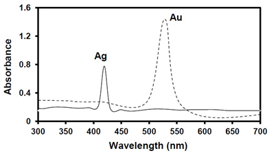
Figure 1: UV-visible spectrum of silver and gold nanoparticles synthesized using Aloe vera gel extract.
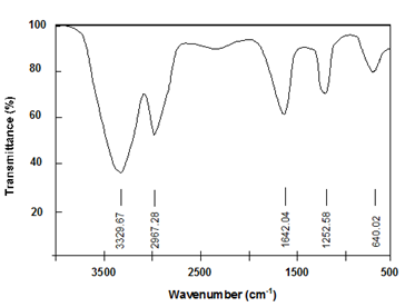
Figure 2: FTIR spectrum of Aloe vera gel extract.
The morphology of silver and gold nanoparticles was found to be poly-dispersed spherical particles, when analyzed using transmission electron microscope. The size of silver and gold nanoparticles ranged in size between 12 - 40 nm and less than 15 nm respectively (Figures 3 and 4). The XRD analysis of AgNPs and AuNPs through bio-reduction and stabilization of Aloe vera showed a Bragg reflections peak at (111), (200), (220) and (311) confirming face centered cubic lattice structure of the formed nanoparticles (Figures 5 and 6).
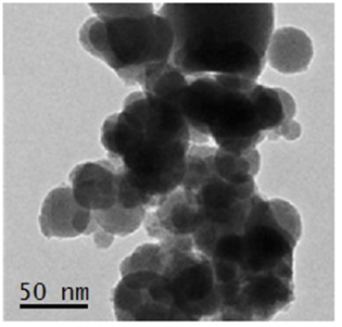
Figure 3: TEM images of silver nanoparticles derived by reduction and stabilization of Aloe vera gel extract.
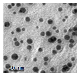
Figure 4: TEM images of gold nanoparticles derived by reduction and stabilization of Aloe vera gel extract.
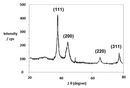
Figure 5: XRD peaks of silver nanoparticles derived by reduction and stabilization of Aloe vera gel extract.
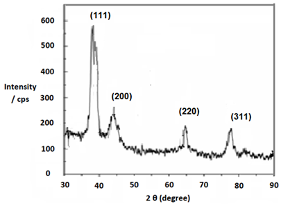
Figure 6: XRD images of gold nanoparticles derived by reduction and stabilization of Aloe vera gel extract.
3.2 Antibacterial activity of nanoparticles
The antimicrobial effect of the synthesized nanoparticles were investigated by measuring the zone of inhibition using varying concentration of 10, 20, 30 and 40 µl/ml, after incubating for 24 h at 35°C. The results showed that both silver and gold nanoparticles exhibited greater inhibition zone against Staphylococcus aureus (Gram positive) followed by Escherichia coli (Gram negative) bacterium at 30 µl/ml (Table 1). Several researchers had proposed corroborated mechanisms for the inhibition of microbial growth due to nanoparticles. The inhibition of bacterial growth depends on the cell wall and its protein, which in turn determines the affinity and penetrating capability of nanoparticles based on its size and charge it carries [12]. In adsorption mechanism, the nanoparticles adhered on cell wall which increased the permeability of membrane leading to intracellular leakage and subsequently to deactivation of enzyme and denature of DNA molecule leading to cell death. E. coli being gram negative bacteria has thick layer of peptidoglycan compared to S. aureus, thus showed increased resistance [13-15]. While the free radicals mechanism proposed that the bactericidal activity of silver nanopaticles was most likely due to the attachment of the silver nanopaticles to the cell wall [16].
|
Microorganism |
Inhibition Zone (mean ± SD) mm of nanoparticles synthesised from Aloe vera |
|||||||
|
Silver nanoparticles (µl/ml) |
Gold nanoparticles (µl/ml) |
|||||||
|
10 |
20 |
30 |
40 |
10 |
20 |
30 |
40 |
|
|
Staphylococcus aureus |
14.3 (±1.5) |
14.9 (±0.7) |
15.1 (±0.6) |
15.3 (±0.7) |
15.2 (±.0.8) |
16.8 (±0.9) |
17.3 (±1.2) |
17.7 (±1.3) |
|
Escherichia coli |
11.2 (±1.2) |
11.4 (±1.3) |
11.9 (±1.1) |
12.2 (±0.9) |
12.7 (±1.5) |
13.5 (±1.6) |
14.4 (±0.9) |
14.9 (±0.7) |
Table 1: Antimicrobial effect of silver and gold nanoparticles in well diffusion method.
3.3 Evaluation of antimicrobial effect on nanoparticles impregnated cotton fabric
The nanoparticles impregnated cotton fabric was evaluated by parallel streak method by monitoring the bacteria growth retarding effect of fabric. The results of bioassay showed that the bacterial growth of S.aureus and E. coli was completely inhibited in the fabric treated with silver and gold nanoparticles . The mechanisms of antibacterial activity of nanoparticle are due to the large surface area and high surface energy of nanomaterials which leads to high durability and immobilization in the fabric. The nanoparticles impregnated fabric inhibited the metabolism due to the interaction with cell wall bounded sulphur proteins resulting in bactericidal effect [17, 18].
4. Conclusion
In the present study aqueous extract of Aloe vera gel proved as a potential candidate for synthesizing nano sized silver and gold particles. The colour change of synthesizing medium occurred due to excitation of surface plasmom resonance which was confirmed through visual and spectral observation at 420 and 530 nm. The synthesized silver and gold nanoparticles were found to be spherical shaped particles with fcc structure, that exhibited inhibition against E.coli and S.aureus at 30 µl/ml. The nanoparticles impregnated fabric exhibited and inhibitory effect at a dosage of 10 µl/ml of silver and gold nanoparticles against human pathogens E.coli and S.aureus using streak plate method.
Funding
The current research investigation was funded by the University Grant Commission of India under the minor research project – FRMP-6368/16 (UGC-SERO).
Acknowledgements
We gratefully acknowledge UGC for funding the minor research project FRMP-6368/16 (UGC-SERO). We are grateful to the Management of Sir Theagaraya College - Chennai and Hindustan Institute of Technology and Science – Chennai. And we also thank SAIF-IITM for the TEM and XRD analysis.
References
- Hobman JL, Crossman LC. Bacterial antimicrobial metal ion resistance. J Med Microbiol 64 (2015): 471-497.
- Lemire J, Harrison JJ, Turner, RJ. Antimicrobial activity of metals: mechanisms, molecular targets and applications. Nat Rev Microbiol 11 (2013): 371-384.
- Park Y, Hong YN, Weyers A, et al. Polysaccharides and phytochemicals: a natural reservoir for the green synthesis of gold and silver nanoparticles. IET Nanobiotechnol 5 (2011): 69-78.
- Rathor N, Mehta AK, Sharma AK, et al. Acute effect of Aloe vera gel extract on experimental models of pain. Inflammation 35 (2012): 1900-1903.
- Li G, He D, Qian Y, et al. Fungus-mediated green synthesis of silver nanoparticles using Aspergillus terreus. Int. J. Mol Sci 13 (2012): 466-476.
- Subramani K, Shanmugam BK, Rangaraj S, et al. Screening the UV-blocking and antimicrobial properties of herbal nanoparticles prepared from Aloe vera leaves for textile applications. IET Nanobiotechno l12 (2018): 459-465.
- Viswanathan K, Diviya Bharathi B, Chitra K, et al. Studies on antimicrobial and wound healing applications of gauze coated with CHX-Ag bybrid NPs. IET Nanobiotechnol 14 (2020): 14-18.
- Medda S, Hajra A, Dey U, et al. Biosynthesis of silver nanoparticles from Aloe vera leaf extract and antifungal activity against Rhizopus sp. and Aspergillus sp. Applied Nanosci 5 (2015): 875-880.
- AATCC Technical Manual, American Association of Textile Chemists and Colorists (2009).
- Sivakumar T, Rathimeena T, Shankar P. Production of silver nanoparticles synthesis of Couroupita guianensis plant extract against human pathogen and evaluations of antioxidant properties. Int J of Life Sci 3 (2015): 333-340.
- Yusuf ZA, Shemau AA, Haalima SA. Phytochemical analysis of the methanol leaves extract of Paullinia pinnata linn. J. of Pharmacognosy and Phytothera 6 (2014); 10-16.
- Slavin YN, Asnis J, Häfeli UO, et al. Metal nanoparticles: understanding the mechanisms behind antibacterial activity. J Nanobiotechnol 15 (2017): 65.
- Ajitha B, Reddy YAK, Reddy PS. Biogenic nano-scale silver particles by Tephrosia purpurea leaf extract and their inborn antimicrobial activity. Spectrochimica Acta Part A 121 (2014): 164-172.
- Kim TH, Kim M, Park HS, et al. Size dependent cellular toxicity of silver nanoparticles. J Biomed Mater Res A 100 (2012): 1033-1043.
- Hajipour MJ, Fromm KM, Ashkarran AA, et al. Antibacterial properties of nanoparticles. Trends Biotechnol 30 (2012): 499-511.
- Oves M, Aslam M, Rauf MA, et al. Antimicrobial and anticancer activities of silver nanoparticles synthesized from the root hair extract of Phoenix dactylifera. Mater. Sci. Eng. C 89 (2018): 429-443.
- Patra JK, Gouda S. Application of nanotechnology in textile engineering: an overview. J Eng Technol Res 5 (2013): 104-111.
- Prabhu S, Poulose EK. Silver nanoparticles: mechanism of antimicrobial action, synthesis, medical applications, and toxicity effects. Int Nano Lett 2 (2012): 2-10.

 Impact Factor: * 2.9
 Acceptance Rate: 78.36%
  Time to first decision: 10.4 days
  Time from article received to acceptance: 2-3 weeks