Glomus Tumor of the Nail Bed with Local Pain: A Case Report
Pervez Ahsan1*, Kamruzzaman2, Israt Jahan3, Abul Khair4
1Professor and Head, Department of Orthopedic Surgery, Ibn Sina Medical College Hospital, Dhaka, Bangladesh
2Assistant Registrar, Department of Orthopedic Surgery, Ibn Sina Medical College Hospital, Dhaka, Bangladesh
3Clinical Research Assistant, Department of Orthopedic Surgery, Ibn Sina Medical College Hospital, Dhaka, Bangladesh
4Medical Officer, Department of Orthopedic Surgery, Ibn Sina Medical College Hospital, Dhaka, Bangladesh
*Corresponding Author: Pervez Ahsan, Department of Orthopedic Surgery, Ibn Sina Medical College Hospital, Dhaka, Bangladesh
Received: 06 February 2023; Accepted: 21 February 2023; Published: 03 March 2023
Article Information
Citation:
Pervez Ahsan, Kamruzzaman, Israt Jahan, Abul Khair. Glomus Tumor of the Nail Bed with Local Pain: A Case Report. Journal of Orthopedics and Sports Medicine 5 (2023): 84-86.
View / Download Pdf Share at FacebookAbstract
Glomus tumors are rare mesenchymal perivascular tumors that often developed in adult age between 30 to 50 years, near fine peripheral neurovascular structures, most notably finger nail beds. A 34 years old woman presented to OPD with pain in the left little finger nail for last five years. On examination, we found Love’s pin test and Hildreth’s test was positive, cold sensitivity test was equivocal. She was diagnosed as glomus tumor. Surgical intervention used to remove completely the tumor and the outcome was excellent. Histopathology confirmed the diagnosis.
Keywords
<p>Glomus tumors; Finger nail; Hildreth's test</p>
Article Details
1. Introduction
Glomus tumors are rare benign vascular hamartomas that arise from the glomus body [1]. It accounts for about 1-4% of all hand tumors [2]. The tumor can appear at any age, but it is most common between the ages of 30 and 50 [3]. Solitary glomus tumors affect women more frequently than men [4]. Multiple lesions are slightly more likely to occur in men [5]. On average, it takes around seven years from the onset of symptoms to the specific diagnosis [3]. Because of its low prevalence and lack of awareness among primary care physicians, it is frequently misdiagnosed [6,7]. Glomus tumors are clinically characterized by a triad of cold sensitivity, localized tenderness, severe and intermittent pain [8]. There are several clinical tests for glomus tumors, including the Love's pin test (100% sensitivity, 0% specificity, and 78% accuracy), the Hildreth's test (71.4-92% sensitivity, 91-100% specificity, and 78% accuracy), and the cold sensitivity test (100% sensitivity, specificity, and accuracy) [9-11]. The investigation modalities include radiographs, ultrasonography and MRI with contrast enhancement. MRI is the investigation of choice [12]. Surgical excision of the tumor with its capsule is the only option for treatment.
2. Case Report
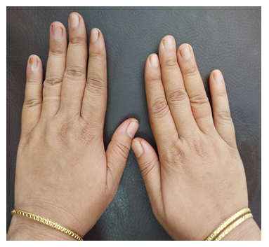
A 34 years old female presented to outpatient department with a five years history of pain in left little finger nail. No history of any preceding trauma. The nail is slightly discolored. She described the incidence of severe pain when exposed to cold. There was no evidence of Raynaud's phenomenon. There was absence of fever, rash or ulcer (Figure 1).

Figure 1: Left little finger showing affected nail.
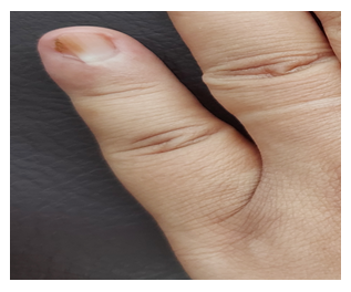
On examination, a sharp localised point of tenderness was found over the nail of the left little finger. Love’s pin test and Hildreth’s test was positive. Cold sensitivity test was equivocal. There was a slight discoloration nail. There was no rise of local temperature, regional lymph node enlargement and the patient was afebrile. The systemic examination revealed normal (Figure 2).

Figure 2: Discoloration of nail bed of left little finger.
3. MRI Findings
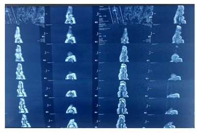
There isointense (to muscle) subcutaneous lobulated soft tissue lesions (0.7 × 0.6 cm) on T1W1, hyperintense on T2W1 and STIR image were seen at dorsal aspect of distal phalanx of left little finger. The lesions exhibited mild heterogeneous enhancement on post contrast T1W1. Thin curvilinear vessels with flow-void in all sequences were seen within the vicinity of the lesions.
All these features were suggested of glomus tumor at dorsal aspect of distal phalanx of left little finger (Figure 3).

Figure 3: MRI showing glomus tumor at distal phalanx of left little finger.
4. Surgical Procedure
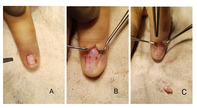
The procedure was carried out under digital block with 1% xylocaine and proximal tourniquet control. A surgical marker was used to mark the location of the tumor on the nail plate. A direct transungual approach was used and proximal nail avulsion was performed to expose the nail bed. A longitudinal incision was made on the nail bed over the tumor. A 0.7 × 0.6 cm shiny, pinkish, encapsulated lesion emerging from the nail bed was discovered. The tumor and its fibrous capsule were carefully dissected (Figure 4). Then we repaired the nail bed. The excised tumor was sent to histopathology, which confirmed the diagnosis.

Figure 4: A,B,C showing the surgical removal of glomus tumour.
5. Discussion
Glomus tumors are rare vascular benign tumors that arise from the neuromyoarterial cells of the normal glomus apparatus of the reticular dermis and are supposed to function in thermal regulation [4]. 50% of these occur under the fingernail between the ages of 30 and 50, and are twice as common in women as in men [13-15]. Three clinical symptoms distinguish glomus tumors: cold sensitivity, localized tenderness, and severe and intermittent pain [8]. Clinical examinations for glomus tumors include the Love's pin test, the Hildreth's test and the cold sensitivity test. There have been several studies on glomus tumor.
According to Salati et al. [16] the patient had an uneventful postoperative period and was symptom free and satisfied at 6 months follow-up.
Jalan et al. [17] in their study found that the postoperative period was uneventful and the patient was asymptomatic with no evidence of recurrence and very satisfied with the cosmetic result as well as with the clinical resolution of her symptoms after 1.5 yrs. follow up.
Ziani et al. [18] at their study conclude that the patient was relieved of functional signs immediately after surgery and the evolution noted no deformation of the affected nail without recurrence of symptoms with a follow-up of 3 years.
Uddin et al. [19] performed surgical excision by direct transungual approach and found that all patients showed dramatic relief of pain after surgical excision.
Suto et al. [20] showed that pain disappeared after surgery, and no recurrence was observed at more than 2 years' follow-up.
In our study, we removed the tumor and its fibrous capsule surgically. The pain was relieved immediately following surgery and the recovery period was uneventful.
6. Conclusion
Glomus tumor is a rare vascular benign tumor. We can diagnose it clinically and radiologically. Surgical excision is the only modality of treatment. After surgical intervention the patient is recovered completely.
References
- Mc Dermott EM, Weiss AP. Glomus tumour. J Hand Surg Am 31 (2006): 1397-1400.
- Mitchell A, Spinner RJ, Ribeiro A, et al. Glomus tumor of digital nerve: case report. J Hand Surg Amx 37 (2006): 1180-1183.
- Kim DH. Glomus Tumor of the Finger Tip and MRI Appearance. Iowa Orthop J 19 (1999): 136-138.
- Chou T, Pan SC, Shieh SJ, et al. Glomus Tumor: Twenty-Year Experience and Literature Review. Ann Plast Surg 76 (2016): S35-40.
- D'Acri AM, Ramos-e-Silva M, Basílio-de-Oliveira C, et al. Multiple glomus tumors: recognition and diagnosis. Skinmed 1 (2002): 94-98.
- McDermott EM, Weiss AP. Glomus tumors. J Hand Surg Am 31 (2006): 1397-1400.
- Fazwi R, Chandran PA, Ahmad TS. Glomus tumour: a retrospective review of 15 years’ experience in a single institution. Malays Orthop J 5 (2011): 8-12.
- Vandevender DK, Daley RA. Benign and malignant vascular tumors of the upper extremity. Hand Clin 11 (1995): 161-179.
- Samaniego E, Crespo A, Sanz A. Key diagnostic features and treatment of subungual glomus tumor. Actas Dermosifiliogr 100 (2009): 875-882.
- Bhaskaranand K, Navadgi BC. Glomus tumour of the hand. J Hand Surg Br 27 (2002): 229-231.
- Giele H. Hildreth's test is a reliable clinical sign for the diagnosis of glomus tumours. J Hand Surg Br 27 (2002): 157-158.
- Matloub HS, Muoneke VN, Prevel CD, et al. Glomus tumor imaging: use of MRI for localization of occult lesions. J Hand Surg 17 ()1992: 472-475.
- Theumann NH, Goettmann S, Le Viet D, et al. Recurrent glomus tumors of fingertips: MR imaging evaluation. Radiology 223 (2002): 143-151.
- Wood W. On painful subcutaneous tubercle. Edinb Med J 8 (1812): 283-291.
- Van Ruyssevelt CE, Vranckx P. Subungual glomus tumor: Emphasis on MR angiography. Am J Roentgenol 182 (2004): 2634.
- Salati SA, Rather A, Khan A. Subungal glomus tumor – a case report. Journal of Pakistan Association of Dermatology (2016).
- Jalan D, Elhence A, Rathore DS, et al. A recurred subungual glomus tumour of the thumb. BMJ Case Rep (2016): bcr2015212963.
- Ziani J, Mernissi FZ, Gallouj S, Monstrous glomus tumor of nail bed clinical and dermoscopic appearance: case report. PAMJ - Clinical Medicine 3 (2020): 60.
- Uddin MM, Biswas SK, Rahman MH, et al. Sub-ungual Glomus Tumor: Study of 20 Cases. Faridpur Medical College Journal 12 (2017): 64-67.
- Suto T, Hardy-Yamada M, Kakinuma T, et al. A case report of a glomus tumour with severe local pain. Pain Clin 19 (2007): 41-42.


 Impact Factor: * 5.3
 Acceptance Rate: 73.64%
  Time to first decision: 10.4 days
  Time from article received to acceptance: 2-3 weeks