Effects of Artemisinin on Peak Sodium Current in Ventricular Myocytes
Huanqiu Song1, Zhuo Ao2, Yuqin Song3, Xiang Li2, Xue Xu1, Cheng Cheng1, Maojing Shi1, Lihua Liu1, Jiatong Wu1, Yuansheng Liu1*, and Dong Han2*
1Department of Emergency, Peking University People’s Hospital, Beijing, China
2National Center for Nanoscience and Technology, Beijing, China
3Zibo Central Hospital, Zibo, Shandong, China
*Corresponding author(s): Yuansheng Liu, Department of Emergency, Peking University People’s Hospital?Beijing, China
Dong Han, National Center for Nanoscience and Technology, Beijing, China
Received: 15 March 2020; Accepted: 24 March 2020; Published: 30 March 2020
Article Information
Citation: Huanqiu Song, Zhuo Ao, Yuqin Song, Xiang Li, Xue Xu, Cheng Cheng, Maojing Shi, Lihua Liu, Jiatong Wu, Yuansheng Liu, and Dong Han. Effects of Artemisinin on Peak Sodium Current in Ventricular Myocytes. Cardiology and Cardiovascular Medicine 4 (2020): 111-117.
View / Download Pdf Share at FacebookAbstract
Background: Previous studies have confirmed that artemisinin can prevent arrhythmia by inhibiting K+ currents. Recent findings have shown that artemisinin attenuates sodium current in nodose ganglion and endocrine cells of rats. This study investigated the effects of artemisinin on peak sodium current in ventricular myocytes.
Methods: Rat ventricular myocytes were isolated by Langendorff reverse aortic perfusion method. Peak sodium current was recorded using the whole-cell patch clamp technique.
Results: The INa was reduced by 50 μM artemisinin, and the steady-state activation and inactivation curves were shifted toward the left. The time constant τ of the steady-state recovery curve increased from 2.89 ms to 7.13 ms.
Conclusions: Artemisinin attenuates INa by modulating the voltage dependence of the Na+ channel similar to the class I anti-arrhythmia agents.
Keywords
Artemisinin; Peak Sodium Current; Antiarrhythmias
Article Details
Introduction
Artemisinin (Art), as an effective antimalarial agent [1], has been widely used for decades. Several other pharmacological effects of artemisinin have been demonstrated, such as cytotoxicity against tumors and cancers [2-3] and antiarrhythmic effects [4]. Previous studies have shown that inhibition of multiple potassium channels, including inwardly rectifying potassium current (Ik1), transient outward potassium current (Ito), and delayed outward rectifier potassium current (Ik) with similar potency, as well as prolongation of action potential duration (APD), are the major mechanisms of the antiarrhythmic effects of artemisinin [5]. A previous study reported that artemisinin affected the amplitude of ionic current in intact nodose ganglion neurons of adult rats by blocking the voltage-gated Na+, K+, and N-type Ca2+ channels?suggesting the probable mechanism of anti-arrhythmia [6]. Recent research has shown the ability of artemisinin to attenuate the voltage-gated Na+ (INa) and delayed-rectifier K+ current (IK(DR)) in endocrine or neuroendocrine cells [7]. All these data show the inhibitory effects of artemisinin on voltage-gated Na+ channels in various cells, but little is known about its effects on ventricular myocytes.
Our previous research confirmed that artemisinin has an antagonistic effect on ventricular arrhythmias induced by increased left ventricular after load [8]. Hence, the present study was undertaken to investigate the effects of artemisinin on peak sodium currents of ventricular myocytes to further understand the mechanism of antiarrhythmic actions of artemisinin.
Abbreviations
APD: Action potential duration
Art: Artemisinin
Ik: Delayed outward rectifier potassium current
Ik1: Inwardly rectifying potassium current
INa: Peak sodium current
Ito: Transient outward potassium current
Materials and methods
Solution The standard Tyrode’s solution (in mM): NaCl 135, KCl 5.4, HEPES 10, NaH2PO4 0.33, MgCl2 1, CaCl2 1.8, Glucose 10, pH adjusted to 7.4 with NaOH. KB solution(in mM): KCl 40, HEPES 10, EGTA 0.5, MgCl2 3, Glucose 10, taurine 20, L-Glutamic acid 50, KH2PO4 20, pH adjusted to 7.4 with KOH. INa Bath solution(in mM): NaCl 20, Choline Chloride 115, MgCl2 1, CaCl2 1.8, BaCl2 0.3, CsCl 5.4, Glucose 10, HEPES 10, CdCl2 0.1, pH adjusted to 7.4 with NaOH. INa Pipet solution(in mM): CsCl 120, HEPES 10, EGTA 10, Na2ATP 5, MgCl2 5, CaCl2 1, Glucose 10, pH adjusted to 7.2 with CsOH.
Artemisinin was dissolved with DMSO and diluted with deionized water to prepare concentration of 50 μM. Nifedipine was prepared at a concentration of 10 mM to inhibit the calcium current. All reagents were purchased from Sigma-Aldrich (Beijing, China).
Isolation of the ventricular myocytesThe animal experiments complied with the Guide for Care and Use of Laboratory Animals drafted by the Institutional Medical Ethics Review Board of Peking University People’s Hospital.
The animals used in this study were Eleven to twelve-weeks old male Wistar rats, purchased from Beijing Vital River Laboratory Animal Technology Co., Ltd.. The method of isolation of the ventricular myocytes was described previously [9]. Briefly, heart was quickly removed and cannulated on a Langendorff device and perfused with Tyrode′s solution (36.5?) for 5 min. Then changing to Ca2+ free Tyrode′s solution (standard Tyrode’s solution without CaCl2) for 10 minutes. Next the heart was enzymatically digested for a period of 15- 20 min with a solution containing collagenase II (200-230 U· L-1). Left ventricular tissue was then excised from the softened hearts and minced gently and filtered with mesh filter. The myocytes were incubated in the KB storage medium at 4? temperature.
Whole-cell patch clamp recordingsINa were recorded in a single ventricular myocyte by whole-cell patch clamp in a voltage clamp mode. The resistance of micropipettes with pipette solution were 2-4 MΩ. An HEKA EPC10 USB single amplifier was used to record current with low pass filtered rate at 3kHz and digital rate at 10kHz. The junction potentials and series resistance were electrically compensated and no leakage correction was applied.
Statistical analysesIn order to correct the current error caused by a discrepancy in the cardiomyocyte size, the current was shown as the current density alternatively. All continuous variables were represented by the mean ± SEM. The differences between the two groups were determined by student’s t-test. A p value of <0.05 was considered statistically significant. All analyses were performed by SPSS version 20.0, and curve fitting was performed by GraphPad Prism version 7.0.
Results
Artemisinin decreased INa in ventricular myocytes
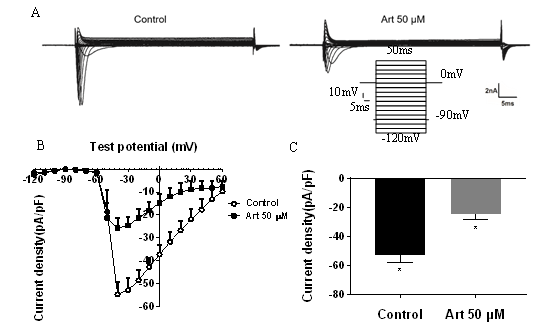
INa was record by a 50 ms depolarization pulse increasing from -120 mV to 50 mV with holding potential at -90 mV. As shown from current traces in (Figure 1A), INa was inhibited significantly with 50 μM artemisinin in the ventricular myocytes. The current- voltage relationship illustrated the effect of artemisinin on INa , especial from -40 mV to 40 mV testing potential (Figure 1B). At −30 mV testing potential, the maximum current density of INa was downregulated from −52.7 ± 5.28 to 24.51 ± 3.85 (n = 5, p < 0.05, Figure 1C).
Figure 1: Effects of artemisinin on INa in ventricular myocytes. (A)current traces of INa in the absence or presence 50 μM artemisinin. (B) current-voltage (I-V) relationship of INa before and after application of 50 μM artemisinin.(C) Summarized data of maximum current density of INa (p < 0.05 vs. control).
The voltage dependence of sodium channel was shifted more negatively by artemisinin
In order to illustrate the inhibitory effects of artemisinin on INa, voltage dependence tests of the Na+ channel were conducted. We analyzed the steady-state activation curve with the same stimulus protocol as mentioned above in current- voltage recording. As shown in Figure 2A, exposure to 50 μM artemisinin, the steady-state activation shifted towards the left. V1/2was decreased from −47.64 mV to −52.12 mV and the slope factor k changed from 2.76 to 2.56. This suggested that 50 μM artemisinin altered the voltage-dependence of the steady-state activation of INa negatively.
We also examined the effect of artemisinin on inactivation kinetics for INa. The steady-state inactivation curve was conducted by a stimulus, which a condition pulse increasing from -120 mV to 0 mV was used to inactivate Na+ channel adequately prior to the testing pulse to 0 mV. In presence of 50 μM artemisinin, the steady-state inactivation curve illustrated in Figure 2B was also shifted negatively. V1/2 was decreased from −78.16 mV to −90.26 mV and the slope factor k changed from 5.37 to 6.77.These results demonstrated that a further negative potential was required for the activation and inactivation of the Na+ channel?resulting in facilitating activation and inactivation at rest potential.
Figure 2: Effects of artemisinin on steady-state activation(A), inactivation(B), and recovery curves(C) of Na+ channel. V1/2 was reduced from −47.64 mV and −78.16 mV to −52.12 mV and −90.26 mV, respectively, and the slope factor k changed from 2.76 and 5.37 to 2.56 and 6.77, respectively. Time constant τ was increased from 2.89 ms to 7.13 ms.
Artemisinin delayed recovery of sodium channel
The steady-state recovery curve was conducted by a condition pulse to -10 mV with holding potential at -120 mV, following a testing pulse to -10 mV with a time interval increasing by 1 ms. As illustrated in Figure 2C, the steady-state recovery curve became less sharp with 50 μM artemisinin and the time constant τwas increased from 2.89 ms to 7.13 ms which indicated that Na+ channel recovered slower from inactivation by 50 μM artemisinin.
Discussion
In the present study, we found that artemisinin inhibited INa in ventricular myocytes and changed the voltage dependence and recovery of sodium channel.
Artemisinin attenuating INa shows similar action to class I antiarrhythmic agents, which decreasing the automaticity and conduction [10]. Three states of Na+ channel (rest, activated, and inactivated) vary in their affinity to the class I antiarrhythmic agents, and activation and inactivation states are higher than rest. Further, different agents show a discrepancy in affinity with these three states of Na+ channels. For example, propafenone has the strongest affinity with open channels, while lidocaine and mexiletine mainly bind to inactivated channels. Furthermore, INa blockers have an unlikely dissociation rate which determines use dependence. Lidocaine and mexiletine dissociate quickly from the Na+ channel and shows no blocking effect on INa at a normal heart rate. However, other INa blockers with longer dissociation duration block INa at a non-tachycardic heart rate [11]. It’s exactly the diversity of dynamic characteristics interacting with Na+ channel determines the different functions of INa blockers.
The present study showed that artemisinin promoted a leftwards shift of the steady-state activation and inactivation curves with more negative V1/2. The results indicated that the threshold potential of Na+ channel was close to the resting potential, facilitating opening of Na+ channels, but inactivation of Na+ channel was also promoted with more negative voltage dependence. As a consequence, INa was downregulated by less Na+ channel available at the resting potential. In addition, the recovery time constant τ was increased accompanied by a less sharp slope of the recovery curve indicating that the Na+ channels recovered gently which enhanced their blocking actions in presence of a slower heart rate. By and large, artemisinin may work on all three state channels of Na+ channel, which contributes to the opening of the Na+ channel, but more negative inactivation voltage and slower recovery reduce the number of available channels leading to INa diminution. But, which state of Na+ channel showing stronger affinity with artemisinin hasn’t make out in our study.
Conclusion
Previous research demonstrated the nonselective inhibition on potassium currents might be the antiarrhythmic mechanism of artemisinin. In present study we showed artemisinin inhibited the peak sodium current but the antiarrhythmic effect of decreasing automaticity and conduction we haven't demonstrated. These results show that artemisinin may play both class I and class III antiarrhythmic functions. There we didn’t include the concentration dependence of artemisinin on peak sodium current, so the applicability of artemisinin needs further study to illustrate.
Declaration of Conflicting Interests
The authors declare that there is no conflict of interest.
Acknowledgement
I hereby declare that this piece of work was fully funded
by National Natural Science Foundation of China Nos.11372012 and 81641002.
Author contributions
Participated in research design: Huanqiu Song, Yuansheng Liu and Dong Han
Conducted experiments: Huanqiu Song, Lihua Liu and Jiatong Wu.
Contributed new reagents or analytic tools: Huanqiu Song and Yuansheng Liu
Performed data analysis: All authors
Wrote or contributed to the writing of the manuscript: Huanqiu Song, Yuansheng Liu and Dong Han
References
- Klayman DL. Qinghaosu (artemisinin): an antimalarial drug from China, Science 228 (1985): 49–55.
- Sun WC, Han JX, Yang WY, Deng DA and Yue XF. Antitumor activities of 4 derivatives of artemisic acid and artemisinin B in vitro. Acta Pharmacol Sin 13 (1992): 541–543.
- Efferth T, Dunstan H, Sauerbrey A, Miyachi H and Chitambar CR. The anti-malarial artesunate is also active against cancer, Int J Oncol 8 (2001): 767–773.
- Crespo-Ortiz MP and Wei MQ. Antitumor activity of artemisinin and its derivatives: from a well-known antimalarial agent to a potential anticancer drug, Biomed Biotechnol 2012: 247597.
- Yang BF, Li YR, Xu CQ, Luo DL, Li BX, Wang HZ and Zhuo J. Mechanisms of artemisinin antiarrhythmic action, Chin J Pharm Toxicol 13 (1999): 169-175.
- Qiao G, Li S, Yang B and Li B. Inhibitory effects of artemisinin on voltage-gated ion channels in intact nodose ganglion neurones of adult rats, Basic Clin Pharmacol Toxicol 100 (2007): 217-224.
- Edmund CS, Wu SN, Wu PC, Chen HZ, Yang CJ. Synergistic inhibition of delayed rectifer K+and voltage-gated Na+currents by artemisinin in pituitary tumor (GH3) cells, Cell Physiol Biochem 41 (2017): 2053-2066.
- Xu X, Zhang Q, Song HQ, Ao Z, Li X, Cheng C, Shi MJ, Fu FY, Sun CT, Liu YS and Han D. Effects of artemisinin on ventricular arrhythmias in response to left ventricular afterload increase and microRNA expression profiles in Wistar rats, Peer J 6 (2018): e6110.
- Li GR, Feng J, Shrier A and Nattel S. Contribution of ATP - sensitive potassium channels to the electrophysiological effects of adenosine in guinea pig atrial cells, J Physiol 484 (1995): 629- 642.
- Campbell TJ. Subclassification of Class-I Antiarrhythmic Drugs -Enhanced Relevance after Cast, Cardiovasc Drug Ther 6 (1992): 519-528.
- Grant AO. Mechanisms of action of antiarrhythmic drugs: from ion channel blockage to arrhythmia termination, Pacing Clin Electrophysiol 20 (1997): 432-444.


 Impact Factor: * 5.6
 Acceptance Rate: 74.36%
  Time to first decision: 10.4 days
  Time from article received to acceptance: 2-3 weeks