Conservative Treatment of Nonunion of the Hamulus Ossis Hamati Using Focused Electromagnetic Extracorporeal Shock Wave Therapy (ESWT) and Extracorporeal Magnetotransduction Therapy (EMTT): A Case Report
Prof. Dr. Karsten Knobloch, FACS1,2*, Dr. Andreas Gohritz3, Prof. Dr. Frank Siemers4
1SportPraxis Hannover Professor Knobloch, Hannover, Germany
2SportPraxis Austria, Perchtoldsdorf, Austria
3Plastic, Reconstructive & Aesthetic Surgery, Hand Surgery, Universitätsspital Basel, Switzerland
4Klinik für Plastische und Handchirurgie/Brandverletztenzentrum, BG Klinikum Bergmannstrost Haale/Saale, Germany
*Corresponding Author: Prof. Dr. Karsten Knobloch, SportPraxis Hannover Professor Knobloch, Hannover, Germany.
Received: 24 July 2025; Accepted: 31 July 2025; Published: 07 August 2025
Article Information
Citation: Karsten Knobloch, Andreas Gohritz, Frank Siemers. Conservative Treatment of Nonunion of the Hamulus Ossis Hamati Using Focused Electromagnetic Extracorporeal Shock Wave Therapy (ESWT) and Extracorporeal Magnetotransduction Therapy (EMTT): A Case Report. Journal of Orthopedics and Sports Medicine. 7 (2025): 392-396.
View / Download Pdf Share at FacebookAbstract
Delayed or nonunion of the hamulus ossis hamati is a rare condition, typically managed surgically by either fixation or excision. This case report highlights the successful treatment of a 30 year-old dentist with a nonunion of the basis of the hamulus type Milch 1-3 in his dominant right hand. Bone stimulation was achieved with a combination of focused electromagnetic extracorporeal shock wave therapy (ESWT, Storz Ultra) and extracorporeal magnetotransduction therapy (EMTT, Storz Magnetolith) with five sessions within 2,5 weeks. Clinical and radiological improvement was observed, within six weeks after the first treatment, highlighting the potential of ESWT/EMTT as superior bone stimulators and a conservative alternative to surgery in nonunion situations of the hand.
Keywords
<p>Nonunion; ESWT; EMTT; Shockwave; Hamulus; Tendons; Muscle; Dentistry</p>
Article Details
1. Introduction
The hamulus ossis hamati (hook of the hamate) plays a vital role in wrist biomechanics, serving as origin of hypothenar muscles (flexor digiti minimi and opponens digiti minimi) and as an anchor for the flexor retinaculum. It also influences the function of the ulnar side of the hand. Fractures of the hamulus are uncommon [1], often resulting from repetitive trauma with force generated by tendons and muscles, or direct falls as acute injuries [2]. When left untreated, nonunion may develop in 24-83% [3-5], leading to chronic pain, functional limitations, and decreased grip strength which is especially significant in professions demanding a lot from the hands, like dentistry.
Surgical options, such as internal fixation or excision, are often considered the gold standard for managing symptomatic nonunion of the hamulus [6]. However, these procedures are associated with inherent risks, including persistent pain, decreased grip strength, and post-operative complications [7]. ESWT has emerged as a minimally invasive alternative for bone stimulation in nonunion situations, demonstrating promising results in various types of delayed bone healing in the hand [8-10]. Blood flow of the scaphoid bone is improved by focused ESWT [11].
Extracorporeal magnetotransduction therapy (EMTT) is a novel energy-based therapy that uses ultra-rapid oscillating magnetic fields. Recently, EMTT has been shown to up-regulate genes paramount for osteogenesis in line with accelerated matrix mineralization in bone healing [12].
This report describes the case of a of hamulus pseudarthrosis successfully treated with a combination of ESWT & EMTT.
2. Case Report
2.1 Patient History
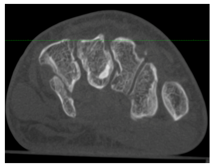
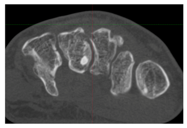
A 30-year-old dentist presented with chronic ulnar-sided wrist pain that had persisted for three months following a bike crash on October 7, 2023 including a fall onto an outstretched dominant right hand. Initial radiographs were unremarkable, but advanced imaging (MRI) taken in December 2023 revealed a nonunion of the hamulus ossis hamati. Conservative management, including immobilization and physical therapy, had failed to alleviate symptoms. On Jan 9, 2024, a high-resolution cone beam CT scan (CBCT, SCS Med Series, Figure 1) was performed, demonstrating a nonunion at the base of the hook type Milch 1-3 [13].
3. Treatment and Outcome
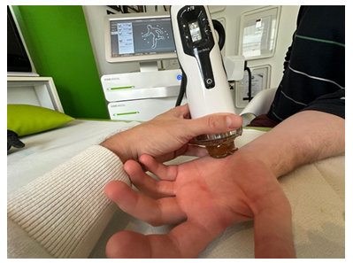
The patient underwent five sessions of focused electromagnetic ESWT (Storz Ultra, Tägerwilen, Switzerland)) and extracorporeal magnetotransduction therapy (EMTT) twice weekly. Focused electromagnetic ESWT was performed from palmar (Figure 2).
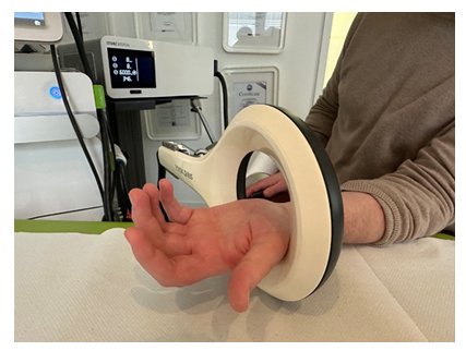
The treatment targeted the site of pseudarthrosis, guided by high resolutation ultrasound with a 22MHz Canon hockey stick probe (Canon Medical Systems, Tokyo, Japan). After focused ESWT, the hand was place within the coil of the EMTT (Figure 3).
The ESWT and EMTT treatment parameters are noted in Table 1.
|
Session |
Focused electromagnetic ESWT (Storz Ultra) |
EMTT (Storz Magnetolith) |
|
1st session |
up to 0,2mJ/mm2, 4000 shots, 18J |
energy 8, 8Hz, 4000 shots |
|
2nd session |
up to 0,4mJ/mm2, 4000 shots, 29,3J |
energy 8, 8Hz, 4000 shots |
|
3rd session |
up to 0,35mJ/mm2, 3000 shots, 30,9J |
energy 8, 8Hz, 4000 shots |
|
4th session |
up to 0,35mJ/mm2, 3000 shots, 33,2J |
energy 8, 8Hz, 4000 shots |
|
5th session |
up to 0,35mJ/mm2, 4000 shots, 45,5J |
energy 8, 8Hz, 4000 shots |
Table 1: Treatment parameters of focused electromagnetic ESWT (energy flux density (mJ/mm2, number of shots, total energy (Joule), Storz Ultra) and EMTT (energy level, frequence, number of shots, Storz Magnetolith) for five consecutive sessions of combined therapy.
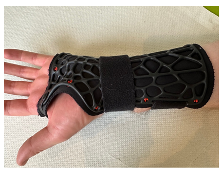
An organic casts was recommended for six weeks during bone stimulation therapy (Figure 4) to foster superior bone healing.
Within six weeks after the initial combined ESWT/EMTT treatment, the patient reported progressive pain relief and improvement in grip strength. Follow-up imaging with high-resolution CBCT scan at six weeks showed evidence of bone healing, and the patient returned to full activity as a dentist without any restrictions or residual symptoms (Figure 5).
4. Discussion
4.1 Nonunion of the Hamulus Ossis Hamati
Pseudarthrosis of the hamulus ossis hamati is uncommon due to its unique anatomical and biomechanical properties. However, when it occurs, it presents significant challenges due to the structure’s small size and limited vascularity. A three-dimensional Chinese study reported that both, the hamate body as well as the hamulus received blood supply from multiple directions and arteries anastomosed extensively outside and inside the hamate [14]. The hamulus receives intraosseus blood supply from palmar, ulnar and the hamulus tip.
Since conservative nonoperative treatment often leads to nonunion situations in more than 50% of the cases, traditional management often involves surgical intervention, such as excision or fixation with screws, both of which carry potential drawbacks. Excision can result in biomechanical changes to the wrist, while fixation may lead to hardware-related complications or incomplete healing.
4.2 The Role of ESWT & EMTT in Delayed Bone Healing
ESWT is a well-established modality for enhancing bone healing by stimulating osteogenesis, angiogenesis, and cellular regeneration [15]. Studies suggest that ESWT is promoting the release of growth factors such as bone morphogenetic proteins (BMPs) and vascular endothelial growth factors (VEGFs) [16-18]. These mechanisms may explain the observed rapid bone stimulation observed in the reported case. In clinical practice, the combination of focused ESWT and EMTT has been successfully used to treat scaphoid [19] nonunion, metacarpal [20] nonunion and humeral [21] nonunions. It appears that mechanotransduction with ESWT and magnetotransduction with EMTT in combination is superior to a single treatment modality. But the combination of ESWT & EMTT is not limited to adults. Even children and adolescents may undergo ESWT & EMTT for bone stimulation without the harm of epiphyseal interactions [22-24].
4.3 Clinical efficacy of ESWT & EMTT for Hamulus nonunion
This case demonstrates an excellent clinical and radiographic outcome following ESWT & EMTT with a fast recovery time. Pain relief, improved grip strength, and functional recovery were consistent findings, aligning with prior studies on ESWT and EMTT for delayed union or nonunion in other anatomical locations. Additionally, ESWT and EMTT provided a non-invasive option that avoided the risks associated with surgical intervention, such as infection, scar formation, or functional deficits which is especially of note in manual-demanding professions like dentistry.
4.4 Advantages of ESWT&EMTT over surgical management
- total noninvasive treatment: ESWT and EMTT require no incisions or implants, thereby reducing the risk of complications.
- Preservation of biomechanics: Unlike excision, ESWT and EMTT maintain the structural and functional integrity of the hamulus ossis hamati.
- Shorter recovery time: Patients undergoing ESWT and EMTT can often resume activities sooner than those undergoing surgery
- Cost-effectiveness: Avoiding surgery reduces hospitalization and overall treatment costs.
5. Conclusion
This case illustrates the potential of combining ESWT & EMTT as a safe, effective, and minimally invasive treatment for nonunion of the hamulus ossis hamati. This modality offers significant benefits, including accelerated healing, pain relief, and preservation of wrist biomechanics, all without the need for surgery. Future studies involving larger cohorts are warranted to establish ESWT & EMTT as a standard bone stimulating conservative therapy for hamulus pseudarthrosis and other small bone injuries.
References
- Plöger MM, Kabir K, Friedrich MJ, et al. Ulnar-Sided Wrist Pain in Sports: TFCC Lesions and Fractures of The Hook of the Hamate Bone as Uncommon Diagnosis. Unfallchirurg 118 (2015): 484-9.
- Suzuki A, Kanda T. Understanding the Injury Mechanism in Hamate Hook Fractures by Investigating Fracture Morphologies: A Case Series Study. Hand (N Y) 20 (2025): 711-719.
- Gardner S, Mudgal C. doi.org/10.1177/17531934114362
- Scheufler O, Andresen R, Radmer S, et al. Hook of hamate fractures: critical evaluation of different therapeutic procedures. Plast Reconstr Surg 115 (2005): 488-97.
- Kadar A, Bishop AT, Suchyta MA, et al. Diagnosis and management of hook of hamate fractures. Journal of Hand Surgery (European Volume) 43 (2017).
- Aykut S, Altun G, Baydar M, et al. Surgical treatment of coronal plane hamate fractures: Clinical and radiological outcomes. Jt Dis Relat Surg 36 (2025): 164-173.
- Sahu MH, Tahir A, Sahu MA, et al. Fractures of the Hamate Bone: A Review of Clinical Presentation, Diagnosis and Management in the United Kingdom. Cureus 16 (2024): e73839.
- Liedl EK, von Schoonhoven J, Prommersberger KJ, et al. Focused High-Energy Extracorporeal Shock Wave Therapy (ESWT) for Bone healing Disorders of the Forearm and the Hand. Handchir Mikrochir Plast Chir 56 (2024): 350-358.
- Quadlbauer S, Pezzei C, Jurkowitz J, et al. Double screw versus angular stable plate fixation of scaphoid waist nonunions in combination with intraoperative extracorporeal shockwave therapy (ESWT). Arch Orthop Trauma Surg 143 (2023): 4565-4574.
- Notarnicola A, Moretti L, Tafuri S, et al. Extracorporeal shockwaves versus surgery in the treatment of pseudoarthrosis of the carpal scaphoid. Ultrasound Med Biol 36 (2010): 1306-13.
- Schleusser S, Song J, Stang FH, et al. Blood Flow in the Scaphoid Is Improved by Focused Extracorporeal Shock Wave Therapy. Clin Orthop Relat Res 478 (2020): 127-135.
- Gerdesmeyer L, Tübel J, Obermeier A, et al. Extracorporeal Magnetotransduction Therapy as a New Form of Electromagnetic Wave Therapy: From Gene Upregulation to Accelerated Matrix Mineralization in Bone Healing. Biomedicines 12 (2024): 2269.
- Milch H: Fracture of the hamate bone. JBJS. 1934, 16:459-62.
- Wang DY, Li X, Shen ZC, et al. Three-dimensional architecture of intraosseus vascular anatomy of the hamate: a micro-computed tomography study. Beijing Da Xue Xue Bao Yi Xue Ban 50 (2018): 245-8.
- Lv F, Li Z, Jing Y, et al. The effects and underlying mechanism of extracorporeal shockwave therapy on fracture healing. Front. Endocrinol 14 (2023).
- Cheng JH, Jhan SW, Chen PC, et al. Enhancement of hyaline cartilage and subchondral bone regeneration in a rat osteochondral defect model through focused extracorporeal shockwave therapy. Bone Joint Res 13 (2024): 342-352.
- Buarque de Gusmao CV, Batista NA, Lemes VTV, et al. Effect of Low-Intensity Pulsed Ultrasound Stimulation, Extracorporeal Shockwaves and Radial Pressure Waves on Akt, BMP-2, ERK-2, FAK and TGF-β1 During Bone Healing in Rat Tibial Defects. Ultrasound Med Biol 45 (2019): 2140-2161.
- Gerdesmeyer L, Zielhardt P, Klüter T, et al. Stimulation of human bone marrow mesenchymal stem cells by electromagnetic transduction therapy - EMTT. Electromagn Biol Med 41 (2022): 304-314.
- Knobloch K. Novel extracorporeal magnetotransduction therapy with Magnetolith and high-energy focused electromagnetic extracorporeal shockwave therapy as bone stimulation therapy for scaphoid nonunion. A case report. Medicine Case Reports and Study Protocols 2 (2021): e0028.
- Knobloch K. Extracorporeal magnetotransduction therapy (EMTT) and high-energetic focused extracorporeal shockwave therapy (ESWT) as bone stimulation therapy for metacarpal non-union - A case report. Handchir Mikrochir Plast Chir 53 (2021): 82-86.
- Knobloch K. Bone stimulation 4.0-Combination of EMTT and ESWT in humeral nonunion : A case report. Unfallchirurg 125 (2022): 323-326.
- Knobloch K, Saxena A, Schaden W. Combined Electromagnetic and Electrohydraulic Focused ESWT and EMTT for Delayed Calcaneal Union in an Adolescent Parkour Athlete - A Case Report. Open Access J Sports Med 15 (2024): 61-66.
- Shafhak T, Amer MA. Focused extracorporeal shockwave therapy for youth sports-related apophyseal injuries: case series. J Orthop Surg Res 18 (2023): 616.
- Omodani T, Takahashi K. Focused Extracorporeal Shock Wave Therapy for Ischial Apophysitis in Young High-Level Gymnasts. Clin J Sport Med 33 (2023): 110-115.


 Impact Factor: * 5.3
 Acceptance Rate: 73.64%
  Time to first decision: 10.4 days
  Time from article received to acceptance: 2-3 weeks