Evaluation of Functional Outcome of Titanium Elastic Nailing in Paediatric Forearm Fracture
Dr. Mohammad Asaduzzaman1*, Dr. Quazi Shahid-ul Alam2
1Assistant Professor, Pediatric Orthopaedic Surgery, Sylhet MAG Osmani Medical College, Sylhet, Bangladesh
2Associate Professor, Pediatric Orthopaedic Surgery, Sir Salimullah Medical College, Dhaka, Bangladesh
*Corresponding Author: Dr. Mohammad Asaduzzaman, Assistant Professor, Paediatric Orthopaedic Surgery, Sylhet MAG Osmani Medical College, Sylhet, Bangladesh.
Received: 07 July 2025; Accepted: 16 July 2025; Published: 25 July 2025
Article Information
Citation: Mohammad Asaduzzaman, Quazi Shahidul Alam. Evaluation of Functional Outcome of Titanium Elastic Nailing in Paediatric Forearm Fracture. Journal of Orthopedics and Sports Medicine. 7 (2025): 360-365.
View / Download Pdf Share at FacebookAbstract
Background: Paediatric diaphyseal forearm fractures are common injuries that are routinely treated with operative stabilisation if they are unstable or displace post-operatively. Titanium Elastic Nailing System (TENS) is an appropriate minimally invasive technique providing stable fixation with maintenance of fracture biology.
Aim of the study: The aim of this study was to evaluate the functional outcome of titanium elastic nailing in Paediatric forearm fracture.
Methods: This prospective observational study was conducted at Sylhet MAG Osmani Medical College Hospital between April 2024 and March 2025. Total 22 paediatric patients presenting with diaphyseal fractures of the forearm were included in this study.
Result: The mean age was 10.5 ± 3.8 years with male preponderance (63.6%). Both bones were fractured in 72.7% of cases. The majority of the fractures healed in 6–8 weeks (68.2%), and all healed at 3 months. Excellent results were seen in 77.3%, good in 18.2%, and fair in 4.5% at final follow-up without poor results. Mean loss of pronation was 6.8 ± 3.2°, supination 8.4 ± 4.1°, and total rotation loss 15.2 ± 5.3°. Complications were rare, with entry site irritation in 9.1% and nail migration in 4.5%.
Conclusion: Titanium elastic nailing is a successful and safe surgical procedure for paediatric diaphyseal forearm fractures, with good functional results and minimal complications.
Keywords
<p>Functional outcome; Titanium elastic nailing; Paediatric; Forearm fracture</p>
Article Details
1. Introduction
Paediatric forearm fractures are one of the most common paediatric injuries in children and account for 30-50% of all paediatric fractures with a predominance of distal radius and ulna [1]. The peak incidence occurs between the ages of 5-14 years, particularly in the context of boys, owing mainly to greater outdoor play, sports-related injuries, and falls from a height during this age of vigorous bone development and high-energy play [1]. Anatomically, the ulna and radius co-operate to allow pronation and supination because of their parallel axis of rotation and the integrity of interosseous membrane [2]. Any loss of alignment after malunion of the fracture, particularly angulation greater than 10 degrees in an older child, has the potential to lead to significant limitation of rotation of the forearm, impacting activities of daily living such as independence, writing, and eating [2,3]. Three-dimensional imaging scans have made rotational malunion and bony impingement the major causes of residual pronation-supination deficits after fracture union, and this shows the importance of anatomical reduction in treating or preventing these injuries effectively [3]. Closed reduction with casting has traditionally been the preferred method of treatment for forearm fractures in children, making the best use of the remarkable remodelling capacity of children [4]. However, this method has shown failure rates of up to 39% in elderly children with unstable fractures, especially the midshaft or proximal third due to the asymmetrical forces exerted by muscles on the fracture pieces [5]. Failed closed reduction, and subsequent re-displacement, angulation, and thus malunion, are at high risk of leading to permanent functional impairment, and this has meant a paradigm shift to surgical fixation of unstable fractures [6]. Loss of reduction in spite of acceptable initial alignment has been observed to happen frequently, which necessitates reassessment of the treatment protocols in order to enhance outcomes. The Titanium Elastic Nailing System (TENS) is now regarded as the gold standard treatment for paediatric diaphyseal forearm fractures, particularly those which are unstable, angulated, or have failed closed reduction [7]. TENS functions under the principle of three-point fixation, in which pre-curved flexible titanium nails are inserted intramedullary to make contact at three points along the medullary canal, provides axial and rotational stability with preservation of the fracture biology [8]. The biomechanical advantage of biological elasticity of the nail allows for controlled micromotion at the fracture site to facilitate external callus and physiological healing under non-rigid immobilization [9,10]. Previous outcome studies on children's forearm fractures using TENS have all uniformly documented union rates near 100%, and its efficacy and reliability [6,7]. Complication includes minor ones like entry site irritation, nail migration, or refracture after removal of the implant, all of which have a tendency to be routinely manageable without significant compromise of function [6]. The less invasive character of the procedure, reduced time in surgery, ease of insertion and removal, and advantage of early mobilization, which is critical in the avoidance of joint stiffness in children, have all helped TENS to become a standard modality used for the treatment of these fractures [7]. In light of the extensive volume of literature supporting the use of TENS, there has been an absence of regional studies comparing functional outcomes in children patients in South Asian populations. This points towards the need for institution-based outcome studies to create contextual evidence-based protocols. Therefore, the present study was undertaken to evaluate the functional outcome of Titanium Elastic Nailing in fractures of the forearm in children.
2. Objective
To evaluate the functional outcome of titanium elastic nailing in Paediatric forearm fracture.
3. Methodology and Materials
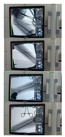
This prospective observational study was conducted at the Department of Paediatric Orthopaedic Surgery, Sylhet MAG Osmani Medical College Hospital, Sylhet, Bangladesh, from April 2024 to March 2025. Total 22 paediatric patients aged between 5 to 15 years presenting with diaphyseal fractures of the forearm were included in this study. Inclusion criteria comprised unstable, angulated, or re-displaced fractures requiring surgical intervention, with informed consent obtained from guardians. Patients with open fractures (Gustilo-Anderson Grade II and above), pathological fractures, associated neurovascular injuries needing repair, or unfit for general anaesthesia were excluded. Preoperative evaluation involved thorough clinical examination and radiographic assessment in anteroposterior and lateral views to determine fracture type, level, and displacement. All surgeries were performed under general anaesthesia with prophylactic antibiotics. Closed reduction was attempted first, followed by Titanium Elastic Nailing (TENS) using pre-bent nails. Nails were inserted via proximal or distal metaphyseal entry points under fluoroscopic guidance, and a below-elbow slab was applied postoperatively for two weeks. Follow-up assessments were performed at 2 weeks, at 6 weeks and at 3 months for suture removal and early complications, early range of motion exercises, clinical and radiological union evaluation, and for final functional outcome assessment, pain and range of motion of supination and pronation as per scale developed by Price CT et al. [11] (Table 1).
|
Outcomes |
Symptoms |
Loss of Forearm Rotation |
|
Excellent |
No complaints with strenuous activity |
<15° |
|
Good |
Mild complaints with strenuous activity |
15°–30° |
|
Fair |
Mild complaints with daily activities |
31°–90° |
|
Poor |
All other results |
>90° |
Table 1: Criteria for evaluation of result Price CT et al. [11].
Radiological union was defined by bridging callus in at least three of four cortices. Data were recorded systematically and analysed descriptively using SPSS version 26. Ethical clearance was obtained from the Institutional Review Board prior to study commencement.
4. Result
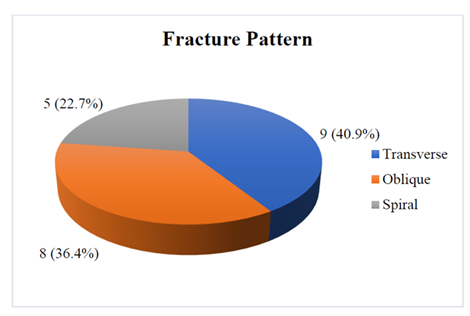
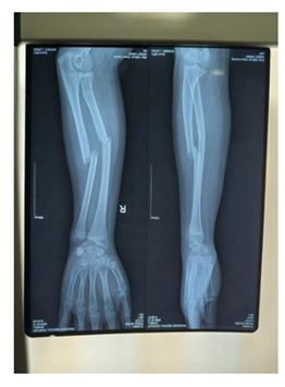
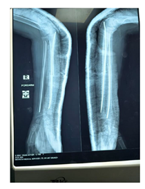
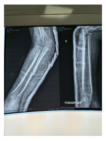
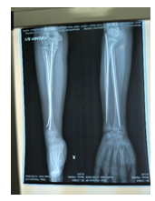
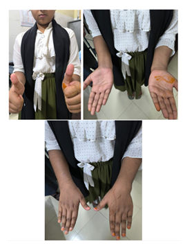
Table 2 highlights the baseline characteristics of the study patients. The mean age was 10.5 ± 3.8 years, ranging from 5 to 15 years. The highest number of patients belonged to the 8–10 year age group (36.4%), followed by 11–13 years (27.3%), 5–7 years (22.7%), and 14–15 years (13.6%). In terms of sex distribution, male patients constituted 63.6% (n=14) while females comprised 36.4% (n=8), indicating a clear male predominance in paediatric forearm fractures. Regarding the fracture site, most patients sustained both bone forearm fractures (72.7%), while isolated fractures of the radius and ulna were observed in 18.2% and 9.1% of patients, respectively. Figure 1 demonstrates the fracture pattern analysis of the study patients. Transverse fractures were most frequent (40.9%), followed by oblique (36.4%) and spiral fractures (22.7%). In terms of postoperative outcomes shown in Table 3, the duration to radiological union was less than 6 weeks in 9.1% of patients, 6–8 weeks in 68.2%, and 9–12 weeks in 22.7%. Functional outcome at 3 months based on Price criteria showed that 77.3% achieved excellent outcomes, 18.2% had good outcomes, and 4.5% had fair results; none had poor outcomes. Complications included entry site irritation in 9.1%, nail migration in 4.5%, and delayed union in 4.5%, while 81.8% experienced no complications. Table 4 presents the mean loss of forearm rotation, with pronation loss at 6.8 ± 3.2 degrees, supination loss at 8.4 ± 4.1 degrees, and an overall loss of rotation of 15.2 ± 5.3 degrees. Table 5 shows the comparison of outcome variables across follow-ups. There were statistically significant improvements in pain scores (p<0.001), pronation (p=0.002), supination (p=0.004), and overall range of motion loss (p<0.001) from 2 weeks to 3 months. Radiological union rates improved from 68.2% at 6 weeks to 100% at 3 months (p=0.004). The proportion of patients with excellent Price criteria outcomes also significantly increased from 31.8% at 6 weeks to 77.3% at 3 months (p=0.003) (Figure 2-7).
|
Characteristics |
Number of Patients |
Percentage (%) |
|
Age group (years) |
||
|
5 – 7 |
5 |
22.7 |
|
8 – 10 |
8 |
36.4 |
|
11 – 13 |
6 |
27.3 |
|
14 – 15 |
3 |
13.6 |
|
Mean ± SD |
10.5± 3.8 |
|
|
Sex |
||
|
Male |
14 |
63.6 |
|
Female |
8 |
36.4 |
|
Fracture Site |
||
|
Both bone forearm |
16 |
72.7 |
|
Radius only |
4 |
18.2 |
|
Ulna only |
2 |
9.1 |
Table 2: Baseline characteristics of the study patients (N=22).
|
Outcome variables |
Number of Patients |
Percentage (%) |
|
Duration to Union (weeks) |
||
|
< 6 |
2 |
9.1 |
|
6 – 8 |
15 |
68.2 |
|
9 – 12 |
5 |
22.7 |
|
Functional Outcome based on Price Criteria (At 3rd month) |
||
|
Excellent |
17 |
77.3 |
|
Good |
4 |
18.2 |
|
Fair |
1 |
4.5 |
|
Poor |
0 |
0.0 |
|
Complication |
||
|
Entry site irritation |
2 |
9.1 |
|
Nail migration |
1 |
4.5 |
|
Delayed union |
1 |
4.5 |
|
Refracture post removal |
0 |
0.0 |
|
None |
18 |
81.8 |
Table 3: Post-operative outcomes of the study patients (N=22).
|
Parameter |
Mean ± SD (degrees) |
|
Loss of Pronation |
6.8 ± 3.2 |
|
Loss of Supination |
8.4 ± 4.1 |
|
Overall loss of rotation |
15.2 ± 5.3 |
Table 4: Mean loss of pronation-supination range of motion.
|
Outcome Variable |
2nd Week (Mean ± SD) |
6th Week (Mean ± SD) |
3rd Month (Mean ± SD) |
p-value |
|
Pain Score (VAS) |
3.8 ± 1.2 |
1.6 ± 0.8 |
0.2 ± 0.4 |
<0.001 |
|
Pronation loss (degrees) |
12.5 ± 5.6 |
8.4 ± 3.9 |
6.8 ± 3.2 |
0.002 |
|
Supination loss (degrees) |
14.2 ± 6.1 |
9.8 ± 4.2 |
8.4 ± 4.1 |
0.004 |
|
Range of motion (total loss) |
26.7 ± 8.3 |
18.2 ± 6.3 |
15.2 ± 5.3 |
<0.001 |
|
Radiological union rate (%) |
– |
68.20% |
100% |
0.004 |
|
Price criteria Excellent (%) |
– |
31.80% |
77.30% |
0.003 |
Table 5: Comparison of outcome variables at different follow-ups (N=22).
5. Discussion
This was a prospective observational study assessing the functional result of the Titanium Elastic Nailing System (TENS) for paediatric forearm diaphyseal fractures. The mean age of the patients was 10.5 ± 3.8 years, of which most were 8–10 years (36.4%), as mentioned in the literature reporting a peak age in the 5–14 years for an increased incidence of high-energy outdoor activity during this age [12,13]. Male dominance occurred in our population (63.6%), as Grabala [14] reported, wherein males had 63.5% of paediatric forearm fracture, perhaps on account of differing gender activity levels and risk-taking. In fracture site distribution, both bone forearm fracture accounted for 72.7% fractures, isolated radius fractures at 18.2% and ulna fractures at 9.1%, which aligns with Sinikumpu15 who defined both bone fractures to be most prevalent, followed by radius-only fractures. In pattern of fracture, the most prevalent was transverse fracture (40.9%), followed by oblique (36.4%) and spiral (22.7%) patterns. Similarly, Vopat et al. [12] and Kumar [16] have also reported distributions in the same context of transverse and oblique fractures being more frequent because of direct trauma and bending type of mechanisms, while rotational forces resulted in spiral fracture. The mean duration to radiological union in this study was within 6–8 weeks in 68.2% of patients, and all fractures united by 3 months. This finding is consistent with Kapila et al. [4], in whom the union time was an average of 7.2 weeks, and Bhat et al. [7], in whom most of the fractures united at 6–8 weeks. Similarly, Saseendar et al. [17] reported the healing of all the patients in 3 months, with TENS contributing to the early healing of the fracture. Functional outcomes based on Price criteria were 77.3% excellent, 18.2% good, and 4.5% fair, with no poor outcome noted. Results are consistent with Bakshi et al. [18], who revealed 90% excellent outcomes, and Kapila R et al. [19] revealed 92 patients with excellent outcomes and 8% patients with good outcomes. Kapila et al. [4] also had 92% good or excellent results, highlighting the consistency of TENS in obtaining excellent functional recovery. That there were no poor results in the study is testimony to the safety and efficacy profile of the treatment. Post-operative complications were minimal, with 9.1% having entry site irritation, 4.5% nail migration, 4.5% delayed union, and 81.8% no complications. These outcomes are similar to complication rates in Jain & Mohanachandran6, wherein there was irritation at the entry site in 7%, migration of the nail in 3.5%, and no refracture on removal of the nail. Bhat et al. [7] also experienced minimal complications with irritation at the entry site in 5.2% and no refractures. Low complication rates here, as also in our own study, indicate the safety of TENS in paediatric fracture management of the forearm. At final follow-up, average pronation loss was 6.8 ± 3.2°, supination 8.4 ± 4.1°, and total rotational loss of forearm 15.2 ± 5.3°, which is functionally satisfactory for daily activity. Kapila et al. [4] reported similar results with the average pronation and supination loss being <10° each, while Saseendar et al. [17] reported total rotational loss of ~16°, as in the current study. Bhat et al. [7] had accounted for a minimally larger loss of supination compared to pronation, which one could also notice in our results with the same pattern. In follow-up analysis, improvement was significant. VAS pain score also improved from 3.8 at 2 weeks to 0.2 at 3 months (p<0.001), consistent with Kapila et al. [4] and Soudy et al. [20], which documented prompt relief from pain by 3 months post-operation. The mean loss of pronation also bettered from 12.5° at 2 weeks to 6.8° at 3 months (p=0.002), and the loss of supination from 14.2° to 8.4° (p=0.004). The total rotation loss also reduced from 26.7° at 2 weeks to 15.2° at 3 months (p<0.001), indicating progressive improvement of functional recovery in accordance with Bhat et al. [7] and Saseendar et al. [17]. Radiological union rates were better at 68.2% at 6 weeks versus 100% at 3 months (p=0.004), in congruence with global reports crediting TENS as a reliable means of union at 3 months [6,7]. Good outcomes by Price criteria were better at 31.8% at 6 weeks versus 77.3% at 3 months (p=0.003) with further improvement on rehabilitation. The findings of this research agree with demographic, fracture pattern, union time, minimal complication, and functional outcome literature that confirms TENS as a safe, effective, and reliable surgery procedure in the management of paediatric forearm diaphyseal fractures.
6. Limitations of the study
In our study, there was small sample size and absence of control for comparison. Study population was selected from one center in Sylhet city, so may not represent wider population. The study was conducted at a short period of time.
7. Conclusion and Recommendations
This current study concludes that Titanium Elastic Nailing System (TENS) is a safe, effective, and minimally invasive surgical option for managing paediatric diaphyseal forearm fractures. This study demonstrated excellent functional outcomes with minimal complications, rapid union within 6–8 weeks in most cases, and near-complete recovery of forearm rotation by three months. TENS offers stable fixation with early mobilisation benefits, making it a reliable treatment modality for unstable fractures in children to ensure optimal anatomical alignment and functional restoration.
References
- Caruso G, Caldari E, Sturla FD, et al. Management of pediatric forearm fractures: What is the best therapeutic choice? A narrative review of the literature. J Orthop Traumatol 22 (2021): 1-12.
- Roth KC, Walenkamp MMJ. Factors determining outcome of corrective osteotomy for malunited paediatric forearm fractures: A systematic review and meta-analysis. J Hand Surg Eur 42 (2017): 545-52.
- Colaris JW, Oei SL, Reijman M, et al. Three-dimensional imaging of children with severe limitation of pronation/supination after a both-bone forearm fracture. Arch Orthop Trauma Surg 134 (2014): 243-9.
- Kapila R, Sharma R, Chugh A. Evaluation of clinical outcomes of management of paediatric bone forearm fractures using titanium elastic nailing system: A prospective study of 50 cases. J Clin Diagn Res 10 (2016): RC12-RC15.
- Nair V, Kumar HS, Devarmani S, et al. Closed reduction nailing or open reduction plating in unstable paediatric forearm fractures: A case of paediatric forearm fracture implant failure. Cureus (2024).
- Jain S, Mohanachandran J. Outcomes and complications of titanium elastic nailing for forearm bones fracture in children: Our experience in a district general hospital in the United Kingdom. Acta Orthop Belg 89 (2023): 539-45.
- Bhat SA, Kumar S, Pathak L, et al. Titanium elastic nailing system, an effective way of pediatric forearm fracture management. Trauma Mon (2022).
- Kumar A. Clinical study on management of diaphyseal fractures of femur in children with titanium elastic nailing system (TENS) [dissertation]. ProQuest Dissertations Publishing (2017).
- Girisha KG. Study of outcome of titanium elastic nails fixation in tibial diaphyseal fracture among children [dissertation]. ProQuest Dissertations Publishing (2020).
- Korhonen L. Pediatric forearm fractures with special reference to operatively treated shaft fractures and ulnar styloid process nonunion [thesis]. University of Oulu (2020).
- Price CT, Scott DS, Kurzner ME, et al. Malunited forearm fractures in children. Journal of Pediatric orthopaedics 10 (1990): 705-12.
- Vopat BG, Kane PM, Christino MA, et al. Treatment of diaphyseal forearm fractures in children. Orthop Rev (Pavia) 6 (2014): 5325.
- Baig M. A review of epidemiological distribution of different types of fractures in paediatric age. Cureus (2017).
- Grabala P. Epidemiology of forearm fractures in the population of children and adolescents: Current data from a typical Polish city [thesis] (2015).
- Sinikumpu JJ. Forearm shaft fractures in children [dissertation]. University of Oulu (2013).
- Kumar P. A prospective study of surgical management of diaphyseal both bones forearm fractures in children [dissertation]. ProQuest Dissertations Publishing (2016).
- Saseendar S, Chidambaram R, Senthil K. Functional outcomes of titanium elastic nailing in paediatric forearm fractures. J Orthop Case Rep 14 (2024): 176-80.
- Bakshi P, Aggarwal A, Garg R. Evaluation of outcome of pediatric forearm fractures treated with titanium elastic nailing system. J Res Med Educ Ethics 11 (2021): 84-8.
- Parajuli NP, Shrestha D, Dhoju D, et al. Intramedullary nailing for paediatric diaphyseal forearm bone fracture. Kathmandu Univ Med J (KUMJ) 9 (2011): 198-202.
- Soudy ESE, Attia MES, Mohamed HEA. Surgical management of fracture both bone forearm in pediatric using elastic stable intramedullary nail. Egypt J Hosp Med 87 (2022): 2520-6.


 Impact Factor: * 5.3
 Acceptance Rate: 73.64%
  Time to first decision: 10.4 days
  Time from article received to acceptance: 2-3 weeks