Breast Cancer or even an Artistic Fantasy in the Paintings of Rembrandt and Rubens?
Piotr Kosiorek1,2*, Dorota Elzbieta Kazberuk1, Katarzyna Jarzabek1, Magdalena Joanna Borkowska1
1Maria Sklodowska-Curie Bialystok Oncology Centre, Ogrodowa 12, 15-027, Bialystok, Poland
2Department of Clinical Immunology, Medical University of Bialystok, Jana Kilinskiego 1, 15-089, Bialystok, Poland
*Corresponding Author: Piotr Kosiorek, Maria Sklodowska-Curie Bialystok Oncology Centre, Ogrodowa 12, 15-027, Bialystok, Poland.
Received: 20 January 2023; Accepted: 31 January 2023; Published: 28 February 2023
Article Information
Citation: Piotr Kosiorek, Dorota Elżbieta Kazberuk, Katarzyna Jarząbek, Magdalena Joanna Borkowska. Breast Cancer or even an Artistic Fantasy in the Paintings of Rembrandt and Rubens?. Journal of Cancer Science and Clinical Therapeutics. 7 (2023): 65-70.
View / Download Pdf Share at FacebookAbstract
Are only internists able to diagnose a disease based on the patient’s appearance, gait, and behaviour? There are many cases of presenting somatic diseases in the art of painting compellingly. Still, there are also paintings where the disease, and even more so the oncological one, is hidden. It may not be widely known at the time. The work aims to rehabilitate Rembrandt’s Betszeba, wrongly regarded since the 1970s as an example of breast cancer, and to show Rubens’s canvases, which do not hide the deformations of breast tumors. The analysis of impurities, stains and varnish deformations is becoming more and more precise thanks to modern methods of X-ray spectrophotometry. In some cases, we see that an effect once considered a disease resulted from artifacts of reconstruction or restoration of the image. Conclusions: there is strong evidence that in the case of Rembrandt’s “Bethsheba in the Bath”, David’s wife or Henrikje Stoffels did not have breast cancer. Other paintings made by Rembrandt and Rubens illustrate a disease. It is puzzling that the artists tried to reflect the realistic nature of the human body, regardless of the existence of possible pathology.
Keywords
<p>Breast Cancer; Paintings; Rembrandt; Rubens; Spectrometry</p>
Article Details
1. Introduction
Is the painter’s intention to hide secrets in the ordered paintings? Can painted figures mean something more than they represent? Isn’t the choice of the author of the portraits sometimes to hide, omit, or skillfully manipulate the characters, playing his own game with the recipient, highlighting what is invisible, stigmatizing flaws, and sometimes ennobling himself by creating his own story on the spur of the moment? Painting functions as a dialogue between the artist and the viewer. The person in the photo conveyed the mood (the portrait was an emoticon). The arrangement of the figures on the canvas told a story from life (thematic scenes). Analogies to the Old Testament inspired appropriate behaviour (biblical scenes) and showed the essence of human life (play, dance, celebrations, battles, etc.) and man’s earthly surroundings (landscapes, still life). Pictures and drawings, like a book, develop our imagination, draw from the depths of our subconscious, and illustrate an unknown foreign world. Can a disease be expressed in an image? Yes. Death, old age, infirmity, and disability are good examples. So, did the painter have something to hide by presenting a realistic figure in a way that differed from reality? Did he reveal to us the beginning of the illness in the portrayed person?. The work aims to compare breasts in Rembrandt’s paintings of naked women. I will defend Rembrandt and his image of bathing Bethsheba, captured in the toilet while reading a letter from King David. David’s future wife guessed what her fate would be. The scene from the Old Testament is eloquent. The sadness, the thought, the inevitability that her husband was exposed to certain death. This is a typical message for the viewer. Was it Rembrandt’s intention to accentuate Bathsheba’s left breast? Did his then-lover Hendrikje Stoffels, who was posing for him at the time, have a breast tumor? Was its varnish? After many years it changed colour, and contemporaries interpreted it as a disease. I will not defend Rubens because the master painted body deformities many times, including breast tumors six times. Although I am inclined to the current version that he instead painted the faces of the portrayed persons of his women and, according to the opinion of numerous art historians, did not hide the presence of the disease in the represented persons.
2. Material and Methods
I will discuss two examples of the female breast, hitherto erroneously considered as disease elements, undeniably created with the participation of Rembrandt himself in various circumstances of his work.
2.1 Hendrikje/Bathsheba’s Breast Tumor in the Bath
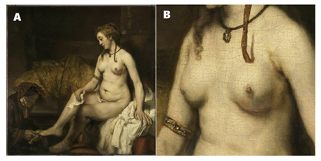
Figure 1A shows “Bethshebe in the bath” (Betshabée ou bain; 1654). Figure 1B is a fragment of this painting depicting the deformity of the left breast. The picture is in the Louvre museum. The thesis presented for over 60 years [1] that Rembrandt’s painted Bathsheba / Hendrikje, David’s wife / and the portrayed partner of David, had a tumor in the left breast (Figures 1A and 1B), becomes outdated [1,2]. More and more data indicate that this is not what the author wanted to tell us about, but it happened by accident [3,4]. A critical study was conducted by Michelle Heijblom et al. in 2012, using a comparison of light colours. She assessed that a breast tumour could not take on such a shade of blue on “dripping” skin. Even mastitis was suspected [5], and the blue colour was associated with Mondor’s disease, i.e. superficial thrombophlebitis [4], but in the case of Mondor’s disease, an adhesion (skin mark) is formed, which in this case excludes this disease, which may be the root cause of breast malignancy. Characteristic skin changes are often seen after breast reconstruction, inflammation, and breast biopsy. Sometimes intense exercise or the pressure of a bra can cause skin folds or fibrous changes on the breast.

Figure 1: Rembrandt painting: Bethesba in bath (A) and fragment presenting breast (B). Source: https://collections.louvre.fr Science iconography courtesy of the Louvre Museum in Paris.
2.1.1 Pettenkofer Conservation: There would be no revelation in discovering the secrets of the conservation of Rembrandt’s paintings if Wolfgang Boehm had not described Max von Pettenkofer’s conservation method popularized in the 19th century [6]. The then innovative (1863!) method of regenerating the “brightening” of the varnish and individual layers of paint of old paintings assumed the restoration of the translucency of the degraded layers of varnish and applied colors. The question posed by Dutch museologists at the turn of the 20th century was to what extent it changed the actual perception and whether there was any mechanical, thermal and chemical damage to the innovative copaiba balm he used, an oleoresin from South America, as a non-polar solvent for dried oil varnishes. In 1990, the ingredients of this balm were identified, and it was found that it contributed to the deformation and dissolution of paints during the “regeneration” of over 150 paintings by various painters, giving them a second life [6]. These phenomena, previously unnoticed, showed an extreme degree of transformation of the painting layer. Interference with the paint concerned: paint deformation (effusion, “mushroom”, undulation) and colour change (stratus, cumulus, cirrus, dot, pustular). The deformation of the varnish resulted in air bubbles and blanching (thermal and physical) (with a wedge gap, a crater, with split grains). These revelations encouraged research using new techniques of thermal-mechanical analysis, e.g. spectrophotometry for individual X-ray layers and images of old masters [6]. Based on the results of numerous conservation works on the painting, the curator of the Louvre museum questioned Rembrandt’s depiction of a breast tumor in Bethsheba.
2.2 Deformity of the Right Breast in an Older Woman Sitting by the Stove
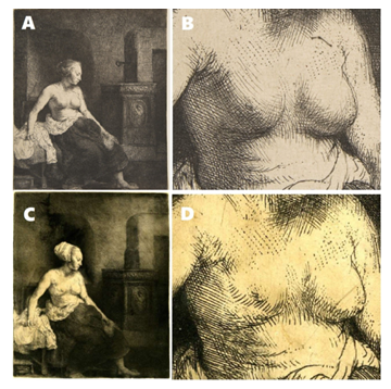
Is this the first recorded evolution of neoplastic lesions? One of the many copperplate prints, an etching from 1658, kept in the Museum in London (Figure 2C and 2D), unlike other prints (Figure 2A and 2B), contains deformations of the right breast previously correctly drawn [7].

Figure 2: Rembrandt etching: Woman Sitting Half-Dressed beside a stove (1658) (A) and fragment presenting breasts (B). Science iconography courtesy of the Metropolitan Museum of Art in New York. Source: https://www.metmuseum.org/art/collection/search/392067 The picture of another sketch: Woman Sitting Half-Dressed beside a stove (1658) (C), and its fragment presenting breast (D) look like the early stage of the first copy of that etching (woman in a bonnet). Science iconography courtesy of the British Museum. Source: https://www.britishmuseum.org/collection/object/P_1848-0911-99
There are at least three hypotheses explaining such differences in etchings [8]. The first hypothesis is that a student of Rembrandt was the author of one or several prints in the early period (page 150) [8]. The second is that the woman in a bonnet was created in 1854 and depicted Henrikje Stoffels after feeding her daughter Cornelia (page 149) [8]. So far, breastfeeding has been described with one breast exposed in paintings. There was a revolution here. To understand this accent of the woman’s two breasts, devoid of sensuality, one has to go back to the beginnings of painting nude nudes from biblical scenes by Rembrandt and his friend Jan Lievens, from the so-called Leiden school in the 1620s and 1630s. It was not until the 1650s in Amsterdam that painting of posing prostitutes began, crossing the borders with moral nudity. Rembrandt enters the canon of imperfect reality. This is reflected in the term aporia.
Moreover, the style of Rembrandt’s sketches of naked women includes the term real-to-life (page 90) [8]. It is a new look at the naked body. Women did not look at the viewer until now, and half-naked or completely naked people did not go beyond the picture’s frame. The engravings are emblems of Rembrandt’s artistic hegemony. The posing woman advertises her nakedness and responds to the viewer’s gaze without hesitation. The painted picture lasts, it is technically finished, but it leaves much room for dialogue and over-interpretation. “The nude as a separate category in art” (page 90) [8]. Figure 2 is undoubtedly one of his first attempts at getting to know a new study of the female nude. A print is like a drawing, a sketch, not fully defined. Did the master notice anything in the breast of the woman portrayed later?. However, there is also a credible indication that Rembrandt may have seen lesions in the older woman’s breast later (Figure 2 A, B). According to David De Witt, the artist knew what he was painting [8]. If Rembrandt drew a prominent breast cancer in an older woman in one of the following sketches of this character [7], why didn’t he depict it in a different, more characteristic and sinister way like in Rubens? However, there are prints of the same drawing without evidence of breast disease in an older woman sitting by the stove. Figure 2. Rembrandt’s etchings from two different periods. Fewer breast-forming lines are visible at first glance (Figure 2D), and the early stage of the NY sketch is immature. Student`s work? (Figure 2C). But other prints in the Rembranthouse in Amsterdam also show no breast distortion. In the dozen surviving sketches, she has no lesions on her breasts [8]. Moreover, it was not the artist’s intention to create these changes [8]. The monograph devoted to Rembrandt’s nudes brings us closer to the circumstances of the creation of paintings and drawings of naked people, but without the context of illness, let alone oncology [8]. De Witt suggests that Rembrandt portrayed the woman for several years (this refers to the woman sitting by the stove without a bonnet on her head), followed by copperplate prints in 1658 showing a study of a woman in a majestic house on Sint Antoniesbreestraat (now Rembrandthuis; Rembrandt’s House). The author must have been aware that he had captured the “evolutions” of breast lesions. “Rembrandt almost certainly saw the compositions for the print directly on the copperplate, not a drawn sketch”[8]. Could the master change the breast image in the engraving so discreetly in a few years? Was it done by his apprentice, paying little attention to the engraving and the layout of the strokes?. Intentions to fully reveal breast cancer do not have to be concealed. Portraits of, among others, breast cancer and other tumors by Lam Qua (the 1830s) are in advanced stages of the disease, known as Trousseau syndrome (venous; thrombotic) can be seen in several paintings of women at Cantona Hospital, in the collection of the missionary doctor Peter Parker at the Yale University Medical Library in New Haven [9].
3. Results
The monograph on the conservation of Rembrandt van Dunn’s paintings mentions many flaws during the long-term restoration of paintings [6]. The use of mass spectrometry [10] and diffusion roentgenography [8] revealed deposits of dirt, dust, varnish, and heavy metal oxides that skillfully concealed the colours of the images. The many years of regenerative phenomena that have taken place so far have significantly contributed to changing the current manifestation of the painting. Amorphous phenomena (e.g. haemorrhages, mushroom-like stains) previously described as chemical reactions of fatty acids with lead, zinc, copper and calcium in pigments, paint additives and paint “dryings”, gave rise to metallic soaps, revealing themselves as aggregates of crystalline or semi-crystalline depending on the chemical composition of the paint. By 1930, most of the 37 Rembrandt or Rembrandt-related paintings in the National Gallery of London, the Dulwich Picture Gallery and the Wallace Collection still had numerous conservation changes, such as cleaning, lining, relining), Varnishing, Revarnishing, Renewing, Pettenkofering and Polishing. In the years after 1950, the primum non-nocere conservation method, i.e. the lining and relining of oil paintings, was dominant, while at the end of the 20th century, the layer kneading method (impasto) was used. Irrespective of the oil painting technique used (glazing, alla prima, impasto), numerous conservation changes and the natural environment for storing paintings did their job.
4. Conclusions
4.1 Where is the Disease? The Breast of Bethsheba and the Three Graces by Rubens
Assuming that the artist’s beloved lady Henrickje Stoffels (Figure 1A) was an extra for portraying Bethsheba (1654), the life span from painting the picture to her death (probably from tuberculosis) was nine years. Therefore, the thesis of the 1980s about a breast tumour that, being visible through the skin layers of the breast, would be in a much-advanced state of cancer with metastases to the lymph nodes is rejected [11]. Inflammation of the breast with the involvement of lymph nodes due to tuberculosis was also considered [3]. But no one expected more research into restoring old Dutch paintings in 2013. X-ray spectrometry’s first examination describing the deformity change on the left breast confirmed the varnish artifact. Conclusion: the oil painting, reconstructed, supplemented and thus “repainted” for many years, significantly changed the image of the female breast [2]. Subsequent studies only confirmed this thesis [4, 8, 10, 12, 13].

Figure 2: Rembrandt etching: Woman Sitting Half-Dressed beside a stove (1658) (A) and fragment presenting breasts (B). Science iconography courtesy of the Metropolitan Museum of Art in New York. Source: https://www.metmuseum.org/art/collection/search/392067 The picture of another sketch: Woman Sitting Half-Dressed beside a stove (1658) (C), and its fragment presenting breast (D) look like the early stage of the first copy of that etching (woman in a bonnet). Science iconography courtesy of the British Museum. Source: https://www.britishmuseum.org/collection/object/P_1848-0911-99
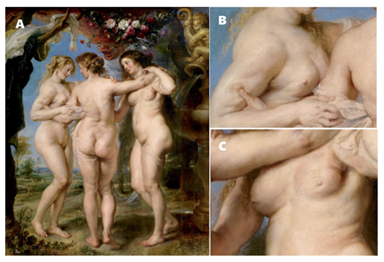
Another well-known breast cancer study painting is The Three Graces (1639) by Rubens [12]. Three women with whom the artist was associated were probably portrayed (Figure 3). Going from the left–Eufrozyne (“joy”) seems to bear the facial features of Rubens’ first wife, Isabella Brant, with whom the painter spent almost twenty years until she died in 1626. Talia (“blossoming”) on the right is, in turn, undeniably a portrait of the artist’s second wife, Helena Fourment, who, as a very young girl, entered the life of an ageing widower. She makes it full of passion and joy that all Rubens’ biographers write about. The third grace (in the middle) is Aglaia (“radiant”), in which Rubens’ biographers see the features of his last lover. In two of them, changes in the breast suggesting a disease process can be discerned (Figure 3B, 3C) [12]. Was it so? Is it just another artefact of many years of reconstruction and renovation of the painting? The lack of published research on the primary reconstruction of the image gives us time for numerous interpretations. Under considerable magnification, we see that the changes in Rubens’ breasts are serial. These include distortion of the breast contour, change in the colour of part of the skin, change in skin texture, retraction of the nipple, and protrusion of axillary lymph nodes. These are undoubtedly features of the breast disease process presented by Rubens [12]. Similar to an isolated, well-circumscribed nodular lesion of the left breast in the painting “Judith with the Head of Holofernes” (1617) [14]. The fact that without coexisting changes is puzzling. Could it be an artefact? Or maybe it’s a captured stage of the onset of the disease. Another painting by Rubens from 1625 of Isabela Brant does not show changes in her breasts. Is Isabela’s similarity with Judith merely coincidental? Maybe someone else posed for the painter? As in the case of other paintings by Rubens, we do not have any explanations or additions about the portrayed people. In “Three Graces” (Figure 3), in the middle portrayed person, one can detect scoliosis and a positive Trendelenburg sign, indicating hip dislocation, nonunion of the femoral neck, coxa valga (valgus hip), lack of hip adductors and abductors function, hip arthritis and sacroiliac [15]. Mild hypermobility syndrome can be suspected in all three (maybe all three were sisters? - Jaccoud’s syndrome/arthropathy and familial joint laxity syndrome, e.g. Ehlers-Danlos?). On the other hand, the left portrayed grace shows signs of hyperlordosis (swan neck), twisted wrist joint, dorsiflexion of the fifth metacarpal bone, and swan finger [15]. Rubens himself suffered from joint changes in the course of rheumatoid disease, and just before his death, he seemed to emphasize this in The Three Graces.
5. Discussion: We see More and More
Nowadays, attention is paid to non-invasive and non-destructive tests of pigments, paints and artistic images using X-ray imaging methods [13]. Among the many X-ray fluorescence methods (XRF) as the primary elemental analysis and imaging method, only combined with X-ray diffraction (XRD) is used. Microscopic XRF (μ-XRF) is a variant of the XRF method capable of visualizing the elemental distribution of primary metals. We detect size traces from 1 μm to 100 μm present in multilayer micro-samples taken from images [13]. The key marker of many of Rembrandt’s works turned out to be the composition of the pigment, or rather a mixture of pigments obtained by the artist by mixing them and applying them to individual critical places of the painting, giving multilayer and spatiality to the set [6]. Thanks to multimedia X-ray diffraction analysis, Rembrandt’s impasto composition containing plumbonacrite was obtained [16]. So another of the secrets of Rembrandt’s painting workshop has been revealed. And all these thanks to the groundbreaking technique of synchrotrons, examining similar pigment compositions from three masterpieces, Portrait of Marten Solemans (1634) from the Rijksmuseum, Bathsheba in her bath (1654) from the Louvre museum and Suzanne (1636) from the Maurithuis museum in The Hague.
6. Summary
“Based on historical texts, we believe that Rembrandt added lead oxide, or litharge, to the oil for this purpose, turning the mixture into a paste-like paint,” noted Marine Cotte, expert and art restorer at the European Synchrotron Lab in Grenoble. The master’s brushstroke is an impasto component – plumbonacrit (a lead compound with the Pb5(CO3)3O(OH)2 structure), while the European Synchroton (ESRF) combined X-ray diffraction of high angular resolution (HR-XRD) with micro-ray diffraction, showing us the places where the characteristic “paste” [16]. Recently, attention has been paid to the increasing amount of impurities accumulated over the centuries on the canvas and canvas, visible in the technique of micro and macro photography and X-ray diffraction scans [6]. The multicultural material of paints, which glow in different wavelengths of light and form flickering micro-crystals under the microscope, is fascinating, but at the time, it was unknown to the painter. In my oncology work, I meet patients after breast reconstruction, with breast cancers that have not yet been operated on, or after their removal (mastectomy). He realizes that comparing the colour of the patient’s skin with the image of changes in the picture [4] in the pathological place does not reflect the changes inside the tissues, even if we compare high-resolution photos of the skin and analyze the skin centimetre by centimetre. As in life, so painting; when we see a picture, we revel in its beauty and lose ourselves in it completely [1]. But we are not interested in the shortcomings of the paint that comes off, cracks in the varnish, the lines of the canvas distortion, or the dirt on the old masters visible to the naked eye [6]. We should pay attention to capturing the moment that the author gave us in the message. Artists create works, and viewers appreciate those years later. Beauty is always in the eye of the beholder, regardless of the theory explaining the imperfections of the image we are looking at.
Declarations
Ethical Approval
The work does not require the participation or consent of humans and animals. It is about refuting the pseudoscientific phenomenon of identifying or naming probable pathologies (breast cancer) in the centuries-old nude paintings by Rembrandt and Rubens. The work aims to show that even the paint's composition or the varnish's reaction to the primer can refute the certainties quoted many times in scientific publications.
Competing Interests
The author has no financial or personal interests or obligations regarding the subject of this work.
Authors Contributions
P.K. and K.J. analyzed the data and drafted the manuscript. D.E.K. and M.J.B. participated in data analysis and extensively reviewed the manuscript. All authors read and agreed to the published version of the manuscript.
Funding
The work does not have any funding.
Availability of Data and Materials
The work is open to the public, on-line analysis of materials made available by museums is free of charge.
References
- Bourne Did Rembrandt's Bathsheba really have breast cancer? Aust N Z J Surg 70 (2000): 231-232.
- Braithwaite PA, Shugg Rembrandt's Bathsheba: the dark shadow of the left breast. Ann R Coll Surg Engl 65 (1983): 337-338.
- Hayakawa S, Masuda H, Nemoto N. Rembrandt's Bathsheba, possible lactation mastitis following unsuccessful Med Hypotheses 66 (2006): 1240-1242.
- Heijblom M, Meijer LM, van Leeuwen TG, et al. Monte Carlo simulations shed light on Bathsheba's suspect breast. J Biophotonics 7 (2014): 323-3
- Zamboni P. The medical enigma of Rembrandt's Bathsheba. J Thromb Haemost 18 (2020): 1268-1270.
- van Dunn E, Noble Rembrandt Conservation Histories. Irma Boom office, Amsterdam, Netherlands (2021).
- Smoluch M, Sobczyk J, Szewczyk I, et al. Mass spectrometry in art conservation-With focus on paintings. Mass Spectrom Rev (2021):
- Greco Rembrandt and breast cancer. Osp Ital Chir 22 (1970): 141-144.
- Grau JJ, Prats M, Diaz-Padron Breast cancer in the Rubens and Rembrandt paints. Med Clin (Barc) 116 (2001): 380-384.
- Nerlich AG, DeWaal JC, Donell ST, et al. Breast cancer in Woman Sitting Half-Dressed beside a stove (1658) by Rembrandt van Breast 64 (2022): 134-135.
- Noorman J, De Witt Rembrandts naakte waarheid. Amsterdam, Netherlands: Museum Het Rembrandthuis (2016).
- Lazzeri D, Lippi D, Castello MF, et al. Breast Mass in a Rubens Rambam Maimonides Med J 7 (2016).
- Dequeker J. Benign familial hypermobility syndrome and Trendelenburg sign in a painting "The Three Graces" by Peter Paul Rubens (1577-1640). Ann Rheum Dis 60 (2001): 894-895.
- Fan BE. Trousseau's Syndrome in 19th Century Qing Dynasty Paintings of Breast Tumors: Early Insights into Cancer and Thrombosis Risk. Semin Thromb Hemost (2022).
- Janssens K, Van der Snickt G, Vanmeert F, et al. Non-Invasive and Non-Destructive Examination of Artistic Pigments, Paints, and Paintings by Means of X-Ray Top Curr Chem (Cham) 374 (2016): 81.
- Gonzalez V, Cotte M, Wallez G, et al. Unraveling the Composition of Rembrandt's Impasto through the Identification of Unusual Plumbonacrite by Multimodal X-ray Diffraction Angew Chem Int Ed Engl 58 (2019): 5619-5622.


 Impact Factor: * 4.1
 Acceptance Rate: 74.74%
  Time to first decision: 10.4 days
  Time from article received to acceptance: 2-3 weeks