Atraumatic Extraction and Immediate Implant Installation
Youginder Singla1*, Rajni Sharma2
1Department of Prosthodontics, Maharaja Ganga Singh Dental College and Research Centre, Sri Ganganagar, Rajasthan, India
2MDS STD, Maharaja Ganga Singh Dental College and Research Centre, Sri Ganganagar, Rajasthan, India
*Corresponding Author: Youginder Singla, Department of Prosthodontics, Maharaja Ganga Singh Dental College and Research Centre, Sri Ganganagar, Rajasthan, India
Received: 02 December 2020; Accepted: 15 December 2020; Published: 28 December 2020
Article Information
Citation:
Youginder Singla, Rajni Sharma. Atraumatic Extraction and Immediate Implant Installation. Journal of Biotechnology and Biomedicine 3 (2020): 111-119.
DOI: 10.26502/jbb.2642-91280032
View / Download Pdf Share at FacebookAbstract
There is an increased resorption of alveolar bone both in horizontal and vertical direction after the extraction of teeth in first 6 months thus affecting aesthetic value of prosthodontics treatment. Implant placement immediately after extraction can decrease resorption of alveolar bone. The clinical study discussed in this article describe the various steps used in atraumatic extraction technique, and then installation of dental implant immediately after extraction. This technique is quiet simple and can be performed easily in clinics and shows excellent results.
Keywords
<p>Atraumatic Extraction, Prostheses and implants</p>
Article Details
1. Introduction
Most common causes of loss of teeth are caries, periodontal and gingival diseases and fracture of teeth. When the teeth are lost especially in anterior region, the apprehension and demand of patient increased dramatically because of asthetic issues. So proper planning is required to meet the requirement of patient and maintain the health of oral tissues [1, 2]. There is increased resorption of the alveolar ridge both in horizontal and vertical direction after the extraction in first 3 months [3, 4]. In anterior teeth, decrease in gingival tissues and lot of aesthetic alterations that hinder the rehabilitation of teeth. There is decreased gingival thickness at margins, change in contour of gingiva and interdental papilla loss with the presence of black spaces [5, 6]. The atraumatic extractions [7], implant placement in socket of extracted tooth [8] is the best alternative to maintain the thickness and color of gingival tissue and is less costly. It also reduces the time of treatment [9] as we have waited for more 3 months to get socket healed and then place the implant. Alveolar bone preservation at the time of tooth extraction, primary stability of the dental implant in alveolar socket in apical direction, the careful reflection of the flap, adaptation of the provisional crown on implant and health of peri-implant tissues are major factors for success of treatment [10, 11]. Maintenance of oral hygiene is a a major contributing factor for the success of immediate implants placement after atraumatic extraction [12]. In this paper a clinical case is presented where the tooth extraction was done using atraumatic extraction kit with immediate implant placement in a mandibular first premolar.
2. Clinical Case Presentation
A 48 year old male patient reported in the clinic for pain in left mandibular 1st premolar. On examination it was found that premolar had horizontal fracture at the level of the marginal gingiva. In X Ray examination, it was found that the premolar had a narrow root canal with little remaining tooth structure making it unsuitable for prosthetic rehabilitation (Figure 1). After thorough analysis and consideration of various option, it was planned extraction of premolar and installation of immediate dental implant. It was verified the systemic condition of the patient and planned atraumatic extraction of the root with ATRAUMATIC EXTRACTION KIT (COWELMEDI) (Figure 2). Atraumatic Extraction Kit is used for the immediate extraction of a tooth with simple procedures according to the type of tooth (e.g., root, apex, and molar) and its position (e.g., mesial and distal). This can also be applied to various cases. A extraction of root is possible by using the rest plate, extraction screw, etc without damaging the Alveolar bone. It is a very fast simple and easy method of tooth extraction as compared to the conventional methods or extraction by the use of periotomes (Figure 3).
- All the coronal structure of tooth is removed by grinding the tooth and is smoothened A hole is created on the tooth to be extracted by using the drill. The Drill should follow the path of the root canal. Root Canal was drilled down to at least 10 mm because extraction is not possible if the screw was not penetrated deep in the root near apex. After connecting the extraction screw to the post driver, it is turned clockwise in order to fix it to the hole that was created. Recommended torque to fix the screw is 30 N/cm. The extraction screw is placed into the hole that was prepared by the extraction drill, and it was fixed to the root. The extraction screw position can be set according to the distal and mesial directions of the adjacent teeth and the position of the tooth to be extracted (Figure 4).
- Torque Head is connected to Extraction screw. After considering the adjacent teeth, extraction screw is inserted into the rest position hole (Figure 5).
- Post driver was connected to extraction screw and then torque wrench was turned in a clockwise direction in order to fix it in the hole.
- The rest plate was connected between the extraction screw and the torque head to protect the adjacent teeth in order to prevent tooth damage. It gives a support to the elevator and torque wrench. - One side of plate is inclined at a 30-degree angle, so that it can provide a support depending on the removal direction. The holes are placed at a 5-mm interval in order to adjust the position of the extraction screw according to the position and distance of the adjacent tooth.
- Then elevator was connected it with the torque head and the tooth was extracted by applying force in a distal or mesial direction.
- Alternatively Torque head can be rotated clockwise by using torque wrench to extract the root (Figure 7).
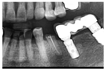
Figure 1: Pre Operative x Ray.
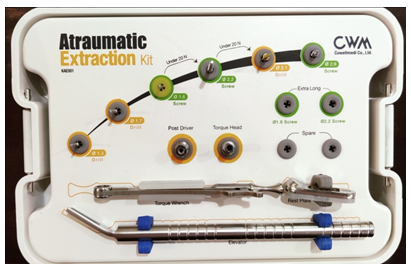
Figure 2: Atraumatic Extraction Kit.
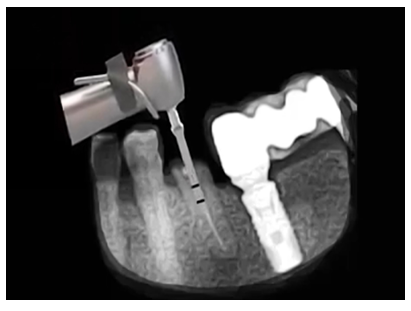
Figure 3: Hole is drilled into root.

Figure 4: Extraction screw is connected to screw driver.
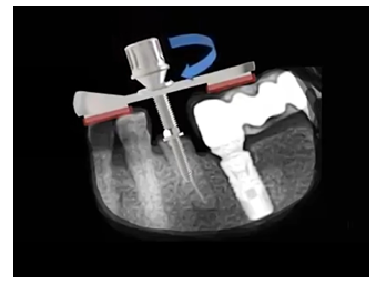
Figure 5: Torque head is placed on the Rest plate.
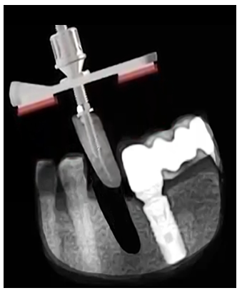
Figure 6: Extraction of root.
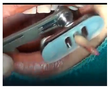
Figure 7: Rotation of Torque head with wrench cause extraction of root Implant installation-COWELMEDI INNO Taper 3.75 x 11.5.

Figure 8: Implant installation – COWELMEDI INNO Taper 3.75 x 11.5.
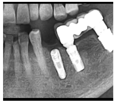
Figure 9: Post operative X Ray.
3. Discussion
The atraumatic extraction is a surgical technique that can present major clinical advantages in the final outcome of prosthetic rehabilitation. It provides greater preservation of alveolar bone and adjacent soft tissue [6, 7]. This technique reduces the chances of loss of thickness and contour of gingival tissues and, therefore satisfactory aesthetics can be achieved. This method can help in the preservation of the alveolar bone. Various techniques have been proposed for this purpose [8, 13-17] with the use of Atrumatic extraction kit is a method which allows in a simple way and with a minimum of trauma to extract the tooth while maintaining the integrity of alveolar bone. The atraumatic extraction may be done when there is fracture of tooth at gingival level and especially when there is a thin bone tissue around root. In the same way, implant installation is done immediately after removal of root to avoid resorption and breakdown of bone after extraction [11-18], and decrease treatment [19] time. The prognosis of the implanted tooth, the causes of loss of tooth, width and depth of alveolar bone beyond the area to be implanted, should be considered before using this technique. If immediate implant placement is done in aesthetic areas, there should be a minimum of 5 mm distance from alveolar crest to contact point for papillae that fill the interproximal space [19]. Thus, to have successful treatment of atraumatic extraction and immediate implant installation, there should be proper case selection, surgical planning and planning of prosthodontics rehabilitation. Postoperative care [2] off course should not be neglected.
4. Conclusion
From presentation of this case and after considering the review of literature, it is concluded that if there is proper case selection and surgical planning then atraumatic extraction with immediate installation of implant is best treatment option for replacement of broken tooth, root or carious tooth. This method can help in the preservation of the alveolar bone.
References
- Suprakash B, Ahammed AR, Thareja A, et al. Knowledge and attitude of patients toward dental implants as an option forreplacement of missing teeth. J Contemp Dent Pract 14 (2013): 115-118.
- Becker W, Goldstein M. Immediate implant placement: treatment planning and surgical steps for successful outcome. Periodontol 47 (2008): 79-89.
- Mummidi B, Rao ChH, Prasanna AL, et al. Esthetic dentistry in patients with bilaterally missing maxillary lateral incisors: a multidisciplinary case report. J Contemp Dent Pract 14 (2013): 348-354.
- Leblebicioglu B, Salas M, Ort Y, et al. Determinants of alveolar ridge preservation differ by anatomic location. J Clin Periodontol 40 (2013): 387-395.
- Horowitz R, Holtzclaw D, Rosen PS. A review on alveolar ridge preservation following tooth extraction. J Evid Based Dent Pract 12 (2012): 149-160.
- Kubilius M, Kubilius R, Gleiznys A. The preservation of alveolar bone ridge during tooth extraction. Stomatologija 14 (2012): 3-11.
- Noelken R, Neffe BA, Kunkel M, et al. Maintenance of marginal bone support and soft tissue esthetics at immediately provisionalized OsseoSpeed‚ NC implants placed into extraction sites: 2-year results. Clin Oral Implants Res 25 (2014): 214-220.
- Muska E, Walter C, Knight A, et al. Atraumatic vertical tooth extraction: a proof of principle clinical study of a novel system. Oral Surg Oral Med Oral Pathol Oral Radiol 116 (2013): 303-310.
- Park JB. Immediate placement of dental implants into fresh extraction socket in the maxillary anterior region: a case report. J Oral Implantol 36 (2010): 153-157.
- Turkyilmaz I, Suarez JC, Company AM. Immediate implant placement and provisional crown fabrication after a minimally invasive extraction of a peg-shaped maxillary lateral incisor: a clinical report. J Contemp Dent Pract 10 (2009): 73-80.
- Kolinski ML, Cherry JE, McAllister BS, et al. Evaluation of a Variable-Thread Tapered Implant in Extraction Sites With Immediate Temporization: A 3-Year Multi-Center Clinical Study. J Periodontol 85 (2014): 386-394.
- De Rouck T, Collys K, Wyn I, et al. Instant provisionalization of immediate single-tooth implants is essential to optimize esthetic treatment outcome. Clin Oral Implants Res 20 (2009): 566-570.
- Botticelli D, Berglundh T, Lindhe J. Hard-tissue alterations following immediate implant placement in extraction sites. J Clin Periodontol 31 (2004): 820-828.
- Oghli AA, Steveling H. Ridge preservation following tooth extraction: a comparison between atraumatic extraction and socket seal surgery. Quintessence Int 41 (2010): 605-609.
- Meneses DR. Exodontia Atraumatica e Previsibilidade em Reabilitacao Oral com Implantes Osseointegraveis - Relato de Casos clinicos Aplicando o Sistema Brasileiro de Exodontia Atraumática Xt Lifting® Rev Port Estomatol Cir Maxilofac 50 (2009): 11-17.
- Mahesh L, Narayan TV, Bali P, et al. Socket preservation with alloplast: discussion and a descriptive case. J Contemp Dent Pract 13 (2012): 934-937.
- Papadimitriou DE, Geminiani A, Zahavi T, et al. Sonosurgery for atraumatic tooth extraction: a clinical report. J Prosthet Dent 108 (2012): 339-343.
- Kan JY, Rungcharassaeng K, Lozada J. Immediate placement and provisionalization of maxillary anterior single implants: 1-year prospective study. Int J Oral Maxillofac Implants 18 (2003): 31-39.
- Younis L, Taher A, Abu-Hassan MI, et al. Evaluation of bone healing following immediate and delayed dental implant placement. J Contemp Dent Pract 10 (2009): 35-42.
- Tarnow DP, Magner AW, Fletcher P. The effect of the distance from the contact point to the crest of bone on the presence or absence of the interproximal dental papilla. J Periodontol 63 (1992): 995-996.
- Malo P, Rangert B, Dvarsater L. Immediate function of Branemark implants in the esthetic zone: a retrospective clinical study with 6 months to 4 years of follow-up. Clin Implant Dent Relat Res 2 (2000): 138-146.
- Turkyilmaz I, Suarez JC. An alternative method for flapless implant placement and an immediate provisional crown: a case report. J Contemp Dent Pract 10 (2009): 89-95.


 Impact Factor: * 5.3
 Acceptance Rate: 75.63%
  Time to first decision: 10.4 days
  Time from article received to acceptance: 2-3 weeks