Durotaxis and VEMFtherapy for Non-invasive Recovery of Mobility and Facial Expression in a Major Burn
Maurizio Busoni*,1, Claudio Urbani2, Sara Zecchetto3 and Simona Laura4
1Master of II Degree in Aesthetic Medicine, University of Camerino - Camerino (MC), Italy
2Urban Medical Beauty – Rome, Italy
3University of Trento – Trento, Italy
4Medical Office Dr. Simona Laura – Vallecrosia (IM), Italy
*Corresponding author: Maurizio Busoni, Master of II Degree in Aesthetic Medicine, University of Camerino - Camerino (MC), Italy.
Received: 12 August 2025; Accepted: 19 August 2025; Published: 01 September 2025
Article Information
Citation: Maurizio Busoni, Claudio Urbani, Sara Zecchetto and Simona Laura. Durotaxis and VEMFtherapy for Non-invasive Recovery of Mobility and Facial Expression in a Major Burn. Fortune Journal of Health Sciences. 8 (2025): 828-834.
View / Download Pdf Share at FacebookAbstract
This study is about the treatment of facial burn scars both from an aesthetic and functional point of view such as fibrosis and retractions that limit facial mobility, full facial expressions, breathing and mouth opening therefore, making chewing difficult. Moreover, the scar determines important facial asymmetries. Our objective is to help the patient find a better quality of life. We treated a major burn patient with obvious motor deficits and relevant facial asymmetries by adopting VEMFtherapy and following the durotaxis method to reduce the impact of fibrosis and retractions of facial biological tissues. The aim was to recover better mobility of facial muscles, greater opening of the mouth and consequently, obtaining recovery of the ability of chewing and a restored skin sensitivity. The results obtained on this patient were relevant. The patient drastically reduced fibrosis, recovered facial expressions and an initial level of facial wrinkling. At the same time, the patient recovered full skin sensitivity, reduced the relief of hypertrophic scars and mitigated the colour difference between scars and normal skin. The patient declares herself fully satisfied with the results. Based on the present experience and other studies relating to VEMFtherapy applied on major burns or complex scars, we believe that VEMFtherapy used according to the durotaxis method can be an effective and safe therapy for all those patients who complain of motor deficits and chronic pain resulting from widespread burns, allowing a significant improvement of the quality of life of the patient.
Keywords
<p>VEMFtherapy, Biodermogenesi, Durotaxis, scar, facial burn, major burn, orofacial disorders</p>
Article Details
Introduction
Treating a patient with major burns, including burns on face, is an extremely complex professional responsibility due to the sum of the problems that we are bound to encounter on multiple levels. In addition to the clinical aspect, we should keep in mind that facial burns cause stigma for the patients and reduce their self-esteem [1], social life and quality of life. Therefore, we should encourage them appreciate all the achieved improvements, even if they are marginal, so that they find the right motivation to follow the recommended therapies. We also know that burn scars, especially for major burns, lead to two particularly serious consequences: water loss from the skin tissue and prolonged periods of skin inflammation. Both of these consequences promote the degeneration of scars, which can evolve into hypertrophic or keloid scars, as well as the formation of significant fibrosis and retractions [2]. Furthermore, the consequences of burns cause serious damages to the patient, both aesthetic, such as facial features, skin texture and pigmentation, and functional, with mobility disruption [3].
Very often, fibrosis and facial retractions in turn lead to aesthetic and functional consequences that heavily impact on the patient's quality of life. On facial burns that require reconstruction with autologous transplant, we witness retraction of the nose, with potential impairment of the respiratory tract, and of the perioral area with a gradual reduction in mouth opening, sometimes reduced to less than one centimetre, forcing the patient to eat only liquid food. All of this is combined with thermal damage to the skin microcirculation [4], both blood and lymphatic, which causes a progressive depletion of the extracellular matrix. One aspect that we consider relevant based on our previous experiences, but which has hardly ever been analysed until today, is the incremental loss of facial expression in patients with major burns who have also suffered extensive facial burns [5, 6]. Retractions keep patients from moving normally, compromising their social life and restricting them to express their emotions.
The scientific literature on major burn victims is abundant in terms of initial emergency treatment, maxillofacial surgery and the immediate post-surgery period, as well as a rich bibliography describing the various levels of discomfort experienced by major burn victims, ranging from the stigma caused by scarring, to loss of self-esteem [1] and reduced mobility [7] which causes a less functional autonomy and leads to various levels of dependence on others, to the daily struggle with chronic pain [8] and to the difficulty of reintegrating into everyday life. What we observed to be lacking is the research pointed out to effective therapies for survivors, applicable after the emergency and surgery period. We know that every year, in times of peace, 300.000 people die from burns and at least 11 million patients suffer burns that require treatment and/or surgery [7]; identifying an effective, safe and replicable therapy for these patients would bring important benefits. On one hand, we can improve their quality of life. From a healthcare perspective, we can reduce their effect on the national healthcare system by reducing the costs associated with chronic therapies. From a social perspective, we can encourage patients to reintegrate into the community and, potentially, into the workplace, supporting their economic independence.
Methodology
With this case report, we wish to illustrate our therapeutic approach against to major burns so that it can be successfully replicated in other patients. In the past, the authors had already developed therapeutic approaches for patients with major burns, such as patients whose faces had been completely destroyed by sulfuric acid [9] or patients with 80% of their bodies covered by third-degree burns [10], using the technology and method described below. In this specific case, we describe the therapeutic path of a major burn patient with third-degree burns affecting the face, wrists, hands and glutes. The patient underwent two surgical procedures on the face based on autologous transplants in August and September 2024. The patient came in for her first visit in March 2025, six months after her last surgery, immediately showing how upset she was about her scars by wearing a flesh-coloured elastic cotton mask with openings for her eyes, nostrils, and mouth. Despite the patient's strong personality, we noted a strong lack of confidence in the future and in the possibility of successfully reintegrating into society, as well as a lack of self-awareness, evidenced by her hair, which was always clean but never styled or coloured.
During the visit, we tried to understand her main concerns and final expectations in order to evaluate whether or not to accept the patient. This precaution is not be-cause of the egoism or the cynicism of the authors, but to the awareness that patients sometimes have expectations that are impossible to meet with their therapeutic path. The patient's expectations were significant but clear and, based on our previous experiences, potentially achievable. She wanted her skin to feel soft again and no longer feel stiff and tight, to the point where she could no longer fully move her face. She wanted to be able to open her mouth wider and with better symmetry, as in time she had noticed that it was becoming increasingly difficult to open her mouth and that this was causing increasing asymmetry. She also wanted to eliminate recurring pain and regain sensitivity on her facial skin. As a matter of fact, the patient really had significant hypertrophic scars with obvious retractions on the right side of the face, with a particularly complex fibrotic bridle starting from the forehead and running down parallel to the nose, extending across the cheek until it split in two, with one end pointing towards the outside of the face and the other towards the right corner of the lips.
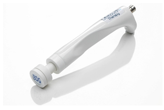
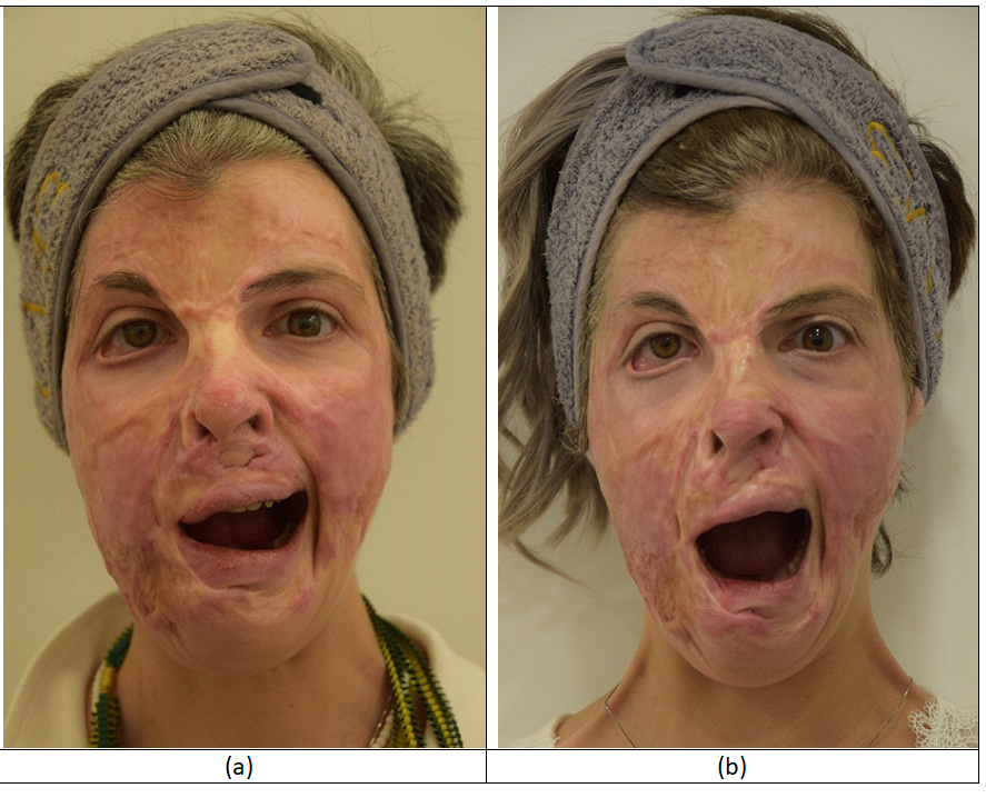
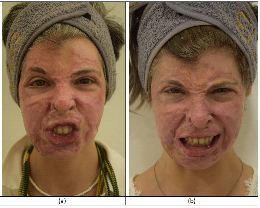
We asked the patient to do facial movements (see figures 3 and 4) which revealed a reduction in facial expressiveness and motility and asymmetrical opening of the oral cavity, again with the most significant deficit on the right side of the face. Before beginning the treatment, the patient signed an informed consent form and a release form, authorising the use of her data and photographs for the purposes of producing this article. In order to meet our patient's expectations, we performed Biodermogenesi therapies, also known as VEMFtherapy, using the Bi-one® LifeTouchTherapy medical device (Expo Italia Srl, Florence, Italy) following two different therapeutic protocols with the specific purposes described below. The therapeutic protocols were developed following the concept of durotaxis [11, 12] aimed at foreseeing the potential future lines of expansion of fibrosis, preventing its formation. The Bi-one® LifeTouchTherapy device offers various therapeutic options based on the use of electromagnetic fields alone or in combination with negative pressure and electrical stimulation waveform. The electromagnetic field is delivered at a variable frequency ranging from 0.5 to 2 MHz with an average intensity of 4 W. Both frequency and intensity are defined by an advanced artificial intelligence system capable of identifying the depth of the damaged tissues and consequently setting up the frequency, while the intensity is defined based on the actual needs of the damaged tissues. The patient underwent an initial cycle of four weekly sessions, using only the electromagnetic field aimed at reducing fibrosis and retractions on her face. The SPECIAL handpiece was used for these sessions (see figure 1).
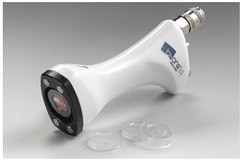
The SPECIAL hand-piece focused on fibrosis and retractions, taking into account the lines of force under-lying the durotaxis method in order to prevent their expansion. Therefore, we treated the scar between the two eyebrows, fibrotic bridle on the right side of the nose, the fibrotic branches on the cheekbones and along the hypothetical nasolabial folds and the perioral area. The action of electromagnetic fields in reducing fibrosis is well known in the literature and was confirmed with the device used in this case history by Marafioti et al. [10], who achieved total remission of a 9 millimeter-thick fibrotic plaque. At the beginning of the fifth session, we used the ACTIVE PLUS FACE handpiece (see figure 2), which allows the combined delivery of electromagnetic fields, negative pressure and electroporation.
This phase lasted for eight weekly sessions, showing significant improvements in the scars, both from an aesthetic and functional point of view. In this phase, we adopted a variable vacuum level ranging from 8 to 10 cent of atmosphere, following the experience of Meirte et al. [13] and Moortgat et al. [14] in the scar therapy with vacuum. At the same time, we distributed a low-intensity electrical stimulation waveform (3.5 VPP on a 500-ohm load) to support the delivery of active ingredients, as demonstrated by Pacini et al. [15]. During this phase, we treated the entire face of the patient with manoeuvres starting from the centre of the face and moving outwards or from the bottom-up. All sessions lasted approximately 20-25 minutes and we asked the patient about her comfort level during the sessions.
Results
At the end of the tenth session, we evaluated the results obtained on the patient thanks to a thorough photographic analysis. The photographs were taken before the start of the treatments, immediately after the end of the tenth treatment and approximately 30 minutes after the end of the tenth treatment.
Figure 3a was taken at T0, while figure 3b was taken immediately after the tenth session. In figure 3b, the tissue still shows a slight redness due to the vacuum. The comparison between the two photographs shows the considerable expansion of the mouth opening obtained thanks to the therapy, contrary to the tendency to progressive restriction reported by the patient before starting the treatment. In addition to the increased motility of the mouth, a reduction in the asymmetry between the two sides is observed which allowed the patient to chew better. Besides that, an increased downward extension of the chin is noted, presumably due to the reduction of retractions that limited facial expression at T0.
Figure 4a was taken at T0, while figure 4b was taken immediately after the tenth session. Also, in figure 4b, the tissue still shows a slight redness due to the vacuum. In the comparison of the two figures, one can observe important improvements due to the reduction of fibrosis and retractions that limited facial expression. Starting from the forehead, it is possible to observe how the patient is once again able to corrugate it clearly. It is noticed how she has recovered the motor control of the right eyebrow aligning it with the left one. At the same time, she can easily tighten the eyelids. The motility of cheekbones and cheeks has definitely improved with a strong symmetry between the two sides and the remodelling of the two nasolabial wrinkles, although not symmetrical, has given a strong facial expressiveness to the patient. The increased mimicry of mouth and lips was particularly observed by the patient.
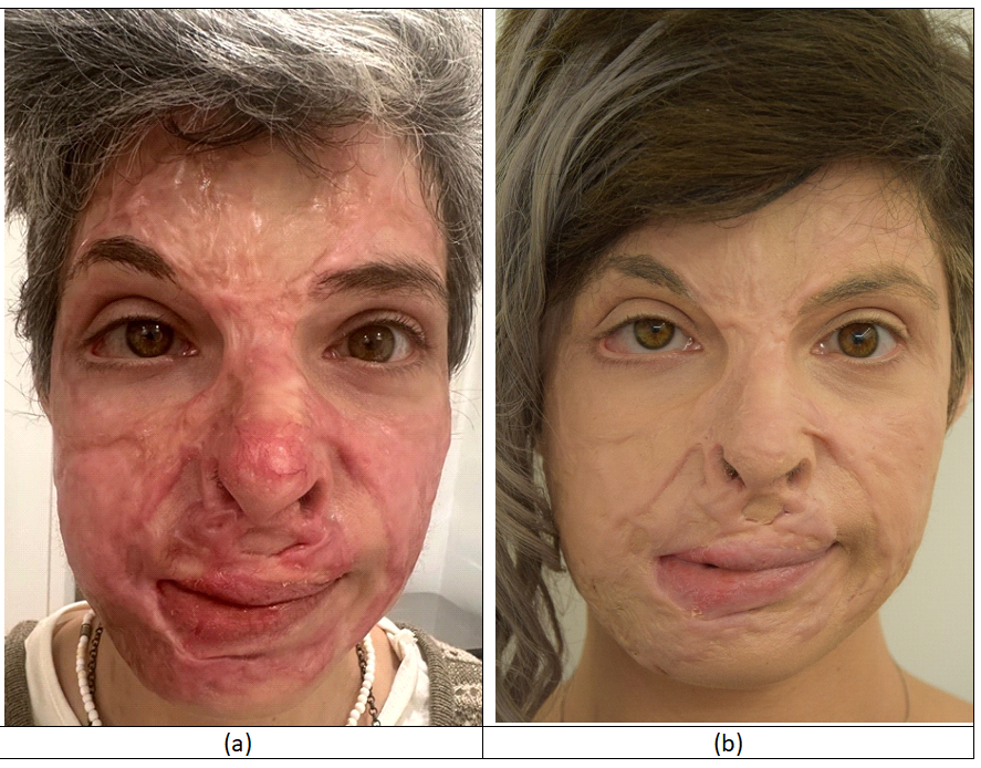
Figure 5a was taken by the patient the week before starting the therapies, while figure 5b was taken about 30 minutes after the end of the tenth session, when the redness caused by the vacuum has normally reabsorbed. In each case, one observes an attenuation of the hypertrophic scar relief that covered the face, a normalization of the skin colour and a greater facial symmetry. It should be noted how the primary fibrotic bridle that starts from the forehead, runs down the right side of the nose and goes down to the cheekbone and the corner of the mouth appears distinctly attenuated. The main limitation of this case history is the lack of ultrasound documentation that could further validate the obtained results. However, we believe that the improvements, both aesthetic and functional, are not debatable. The comparison of figures 5a and 5b shows a total change in the patient’s facial skin, with a reduction of the protrusion of the hypertrophic scars, and hints at a reduction of fibrosis and retractions that allow a better distension of the facial skin.
Even more clearly, figures 3a, 3b, 4a and 4b show that the patient has regained a greater control of her face, again managing to express satisfactory facial expressions, previously inhibited by the significant fibrosis and retractions that characterized the entire face, with particular intensity on the right side, resulting in an evident asymmetry that has been distinctly attenuated thanks to the provided therapies. At the end of the tenth session, the patient stated that the therapies were extremely pleasant and relaxing, giving 10 out of 10 to the comfort level, she declared that she was extremely satisfied with the improvements obtained, both aesthetic and functional, that she chewed very easily, that she no longer felt the recurring pains that affected her and that she found a regained uniform sensitivity of the facial skin which had been partially damaged at T0. There were no side effects in any case, except for a slight redness reabsorbed within 30 minutes due to the action of the vacuum from the fifth to the tenth session. We decided, in agreement with the patient, to schedule several follow-ups to monitor the stabilization of the results and develop a further therapeutic protocol aimed at further standardizing the symmetry of the face and increasing the mobility of the facial tissues.
Discussion
The management of burn patients is one of the most complex issues in medicine, especially when the scars are disfiguring, as in this case of our patient or when they cause disabilities such as chronic pain or reduced mobility which makes it difficult for patients to return to normal life and work, thus limiting their economic independence. In our experience, we have found that some patients build a small and increasingly restricted comfort zone from which they are afraid to come out. They do not want to break a precarious balance they have struggled to build day after day and, above all, they do not want to delude themselves into thinking they can improve. After analysing the results obtained, we believe that many of the previous publications on VEMFtherapy, also known as Biodermogenesi, on electromagnetic fields and vacuum allow to better understand the dynamics. Veronese et al. [9] have already documented how VEMFtherapy led to many of the improvements we observed in a patient whose face had been completely destroyed by sulphuric acid. In that case, the patient recovered full head mobility, which had been damaged by a brutal retraction of the neck whose profile appeared with a linear junction between the chin and the collarbones; also in that case, the patient reported remission of pain and recovery of facial sensitivity. The effect of VEMFtherapy on fibrosis and retractions is documented by Marafioti et al. [10] about a patient with major burns who had fibrosis in her lower limbs that made difficult for her to stand upright. The ultrasound scans show a 9-millimetre-thick fibrotic block on the back of the right leg that completely disrupted the skin and muscle tissue, which completely regressed after a cycle of weekly sessions, so much so that the patient is now able to walk independently without support. The ultrasound scans provided at the end of the sessions also show the restructuring of the skin layers and muscle fibres, justifying the patient's recovered ability to walk. We think that this action also shows itself in the facial muscles, as documented by the increase in mouth opening, better chewing and recovery of facial expressiveness in the patient. This data was also confirmed by Laura et al. [16], who draw attention to the full recovery of finger and hand motility in patients with Dupuytren's Contracture, documenting the reduction in fibrosis causing finger retraction by ultrasound and simultaneously documenting with an infrared camera the recovery of normal vascularization of the affected fingers, which was clearly deficient before treatment. In our opinion, the typical angiogenesis action of the adopted device is particularly important, as demonstrated by Scarano et al. [17] on a group of sixty patients who voluntarily underwent biopsies and histochemical analysis. In the patient under examination, it can be noticed that the burned areas affect the entire area of the facial artery, the inferior labial artery, the horizontal labiomental artery, the angular artery and the infraorbital artery, as well as the supratrochlear vein, the angular vein and the facial vein, particularly on the right side of the face. Impaired microcirculation has always played a significant role in the degeneration of burn scars. So, promoting angiogenesis allows significant volumes of oxygen and nutrients to be returned to the matrix which will be then transferred into the cell membranes thanks to the action of the electromagnetic field which interacts with the Na+/K+ pump. In this specific case, we can imagine that the restoration of the skin capillary network, combined with the stimulation of pre-existing capillaries provided by vacuum [14], allows for a greater exchange with the matrix and consequently with cells and fibroblasts, while the increased activity of Na+/K+ ions through cell membranes leads to a slight increase in skin temperature based on the second law of thermodynamics [18]. These reactions form the basis of Van't Hoff's Law [19] (Nobel Prize for Chemistry in 1901), according to which the biological tissue that is adequately vascularized, rich in oxygen and nutrients, brought to a temperature between 39 and 40°C, multiplies its regenerative capacity fourfold. The stabilization of these temperatures in skin and muscle tissue thanks to electromagnetic fields was demonstrated by Kuraman et al. [20] and later confirmed by Tashiro et al. [21] who observed an increase in temperature of approximately two degrees Celsius involving a depth of treated tissue between 1 and 2 centimetres, stable for the duration of the therapies and for a further 30 minutes after their completion. The thermal effect was further validated by Clijsen et al. [22] in a study that compared patients actually treated with electromagnetic fields and others treated with a placebo. We also consider relevant the reduction in inflammatory conditions typical of both electromagnetic fields documented by Marmotti et al. [23], who found a significant increase in the production of IL-10 cytokines with anti-inflammatory action, and vacuum, as underlined by Marques et al. [24]. We are well aware that a single case report is insufficient to declare a treatment effective and safe, even if supported by other published articles. Precisely for this reason, the authors intend to expand their experience in the treatment of disabling facial scars, collaborating with other colleagues to conduct a multicentre study on a much larger patient base. Our ultimate goal remains to develop a synergy between VEMFtherapy and durotaxis to treat burn-related fibrosis and the related consequences that compromise the quality of life of millions of new patients every year.
Conclusions
When going through the therapeutic path aimed at improving the life of a major burn victim, any obtained clinical results must inevitably be confirmed by an achieved better quality of life of the patient. In this specific case, the obtained benefits were certainly significant from an aesthetic, functional and psychological point of view, so much so that the patient regained the courage to take care of her appearance and improve her self-esteem by comparing herself with other people with greater strength and confidence. In this specific case, VEMFtherapy showed particular effectiveness, safety and pleasantness for the patient. This case history and the other published studies based on the same technology can help burn victims recover a better quality of life.
Author Contributions
M.B. developed the treatment protocols, C.U. and S.L. delivered the therapies, M.B. and S.Z. wrote this article.
Funding
This research received no external funding.
Informed Consent Statement:
Informed consent was obtained from all subjects involved in the study. Written informed consent for publication must be obtained from participating patient who can be identified.
Acknowledgments
Our thanks go to the RigeneraDerma ONLUS Organisation which developed a non-profit project to offer pro bono therapies to women who have been subjected to violence and to people with disabling scars.
Conflicts of Interest
Maurizio Busoni is the member of the Board of Directors of Expo Italia Srl. The other authors have no conflict of interest.
References
- Mehrabi A, Falakdami A, Mollaei A, et al. A systematic review of self-esteem and related factors among burns patients. Ann Med Surg (Lond) 84 (2022): 104811.
- Zhong A, Xu W, Zhao J, et al. S100A8 and S100A9 Are Induced by De-creased Hydration in the Epidermis and Promote Fibroblast Activation and Fibrosis in the Dermis. Am J Pathol 186 (2016): 109-22.
- Burd A. Burns: treatment and outcomes. Semin Plast Surg 24 (2010): 262-80.
- Rose EH. Aesthetic reconstruction of the severely disfigured burned face: a creative strategy for a "natural" appearance using pre-patterned autogenous free flaps. Burns Trauma 3 (2015): 16.
- Peck MD. Epidemiology of burns throughout the world. Part I: Distribution and risk factors. Burns 37 (2011): 1087-100.
- Kilinc DD, Mansiz D. Myofunctional orofacial examination tests: a literature review. BMC Oral Health 23 (2023): 350.
- Dicarla Motta Magnani, Fernanda Chiarion Sassi, Luiz Philipe Molina Vana, et al. Evaluation of oral-motor movements and facialmimic in patients with head and neck burns by apublic service in Brazil. Clinics 70 (2015): 339-345.
- Noma N, Watanabe Y, Shimada A, et al. Effects of cognitive be-havioral therapy on orofacial pain conditions. J Oral Sci 63 (2020): 4-7.
- Veronese S, Brunetti B, Minichino AM, et al. Vacuum and Electromagnetic Fields Treatment to Regenerate a Diffuse Mature Facial Scar Caused by Sulfuric Acid Assault. Bioengineering (Basel) 9 (2022): 799.
- Marafioti S, Veronese S, Pecorella C, et al. Electromagnetic Fields, Electrical Stimulation, and Vacuum Simultaneously Applied for Major Burn Scars. Bioengineering 12 (2025): 179.
- Espina JA, Marchant CL, Barriga EH. Durotaxis: the mechanical control of directed cell migration. FEBS J 289 (2022): 2736-2754.
- Plotnikov SV, Pasapera AM, Sabass B, et al. Force fluctuations within focal adhesions mediate ECM-rigidity sensing to guide directed cell migration. Cell 151 (2012): 1513-27.
- Jill Meirte, Peter Moortgat, Mieke Anthonissen, et al. Short-term effects of vacuum massage on epidermal and dermal thickness and density in burn scars: an ex-perimental study. Burns & Trauma 4 (2016): 27.
- Peter Moortgat, Mieke Anthonissen, Jill Meirte, et al. The physical and physiological effects of vacuum massage on the different skin layers: a current status of the literature. Burns & Trauma 4 (2016): 34.
- Stefania Pacini, Tiziana Punzi, Massimo Gulisano, et al. Transdermal Delivery of Heparin Using Pulsed Current Iontophoresis. Pharmaceutical Research 23 (2006): 114-120.
- Laura S, Giorgio C, Lupo F, et al. VEMF therapy to Treat Dupuytren’s Contracture: A Preliminary Study. Biomed J Sci & Tech Res 60 (2025).
- Scarano A, Sbarbati A, Amore R, et al. A New Treatment for Stretch Marks and Skin Ptosis with Electromagnetic Fields and Negative Pressure: A Clinical and Histological Study. J Cutan Aesthet Surg 14 (2021): 222-228.
- Herbert Kroemer, Charles Kittel, Thermal Physics, 2nd Ed., W. H. Freeman Company (1980).
- https://www.nobelprize.org/prizes/chemistry/1901/summary/
- Kuraman B, Watson T. Thermal build-up, decay and retention responses to local therapeutic application of 448 KHz ca-pacitive resistive monopolar radiofrequency: A prospective randomised crossover study in healthy adults. Int. J. Hyperth 31 (2015): 883-895.
- Tashiro Y, Hasegawa S, Yokota Y, et al. Effect of Capacitive and Resistive electric transfer on haemoglobin saturation and tissue temperature. Int J Hyperthermia 33 (2017): 696-702.
- Clijsen R, Leoni D, Schneebeli A, et al. Does the Application of Tecar Therapy Affect Temperature and Perfusion of Skin and Muscle Microcirculation? A Pilot Feasibility Study on Healthy Subjects. J Altern Complement Med 26 (2020): 147-153.
- Marmotti A, Peretti GM, Mattia S, Pulsed Electromagnetic fields improve tenogenic commitment of Umbialacal cord-derived mesenchymal stem cell; a potential strategy for tendon repair An in vitro study. Stem Cells Int (2018): 9048237.
- Marques MA, Combes M, Roussel B, et al. Impact of a mechanical massage on gene expression profile and lipid mobilization in female gluteofemoral adipose tissue. Obes Facts 4 (2011): 121-9.


 Impact Factor: * 6.2
 Acceptance Rate: 76.33%
  Time to first decision: 10.4 days
  Time from article received to acceptance: 2-3 weeks