Postoperative Bleeding in a Patient with Appendiceal Mucinous Adenocarcinoma and Liver Dysfunction Patient: A Case-Report
Xiaohua Niua#, Chaoming Tanga, Ling Qib, Wenzhong Moa, Haiyang Xina, Mingkun Suna#*
aDepartment of Gastrointestinal Surgery, The Sixth Affiliated Hospital of Guangzhou Medical University, Qingyuan People's Hospital, Qingyuan, Guangdong Province, 511518, China
bDepartment of Clinical Biobank, The Sixth Affiliated Hospital of Guangzhou Medical University, Qingyuan People's Hospital, Qingyuan, Guangdong Province, 511518, China
#First author
Xiaohua Niu
Mingkun Sun
*Corresponding author: Mingkun Sun, Department of Gastrointestinal Surgery, The Sixth Affiliated Hospital of Guangzhou Medical University, Qingyuan People's Hospital, B24 Yinquan South Road, Qingyuan, Guang Dong Province, China
Received: 26 October 2021; Accepted: 03 November 2021; Published: 17 November 2021
Article Information
Citation: Xiaohua Niu, Chaoming Tang, Ling Qi, Wenzhong Mo, Haiyang Xin, Mingkun Sun. Postoperative Bleeding in a Patient with Appendiceal Mucinous Adenocarcinoma and Liver Dysfunction Patient: A Case-Report. Journal of Surgery and Research 4 (2021): 635-648.
View / Download Pdf Share at FacebookAbstract
Background
Appendiceal malignant tumors are rare in the clinic, and the incidence rate of gastrointestinal tumors is only approximately 0.5%. Our aim is to describe our experience with this rare disease and to increase knowledge on the diagnosis and treatment of appendiceal malignant tumors.
Case presentation
We report the case of a 69-year-old woman who was admitted to the hospital due to dyspepsia. The patient was a carrier of hepatitis B virus, and liver dysfunction was diagnosed preoperatively. Abdominal enhanced computed tomography and colonoscopy showed that the appendix was significantly enlarged and dilated, and effusion and appendicitis were observed. Mucinous adenocarcinoma and appendiceal abscesses were not excluded because of the lack of specificity, which makes it difficult to diagnose the disease before a surgery. Laparoscopic appendectomy was performed, and a rapid frozen pathological examination showed a mucinous tumor of the appendix. Intraperitoneal hyperthermic chemotherapy with cisplatin was administered. The patient had abdominal hemorrhage on the fifth day after the surgery. After active treatment, she was discharged from the hospital 19 days after the surgery.
Conclusions
The diagnosis of appendiceal malignant tumors mainly depends on preoperative imaging and microscopic results,and highly suspected patients, rapid pathological examination is needed during the operation., and so on. Notably, for elderly patients with hepatitis B infection and liver dysfunction, there is a probability of postoperative bleeding.
Keywords
<p>Appendiceal mucinous adenocarcinoma, Liver dysfunction, Case report, Hyperthermic intraperitoneal chemotherapy, Postoperative Bleeding</p>
Article Details
1. Background
Appendiceal malignant tumor is a kind of malignant tumor with low incidence in the digestive system, accounting for about 0.5% of gastrointestinal tumors [1], which can be divided into adenocarcinoma and carcinoid. Adenocarcinoma is further divided into mucinous adenocarcinoma, colonic adenocarcinoma, goblet cell carcinoma and signet ring cell carcinoma [2]. Mucinous adenocarcinoma is a histological form of tumor, in which mucous tissue accounts for more than half of the tumor tissue [3]. Primary appendiceal tumors often occur in people over 54 years of age, and more frequently in females than in males [4], but this difference is not significant [5]. At present, there is no conclusive cause of this disease. It is assumed that it is related to the long-term surrounding inflammatory stimulation and inflammatory exudates of the appendix [6]; however, some scholars think that schistosomiasis may lead to the occurrence of appendiceal tumors [7]. Because the disease has no specific manifestation, it is easily misdiagnosed and missed in the clinics, and lacks a standard effective treatment plan. Liver dysfunction refers to liver damage caused by various factors and can be divided into primary and secondary liver dysfunction. Primary liver dysfunction is caused by acute and chronic liver diseases such as hepatitis, liver cirrhosis, liver cancer, etc., whereas secondary liver dysfunction is cause by critical illnesses, severe infections, and surgery [8]. Because of the crucial role of the liver in metabolism, once liver function is impaired, the metabolism and immunity of the body will be affected to varying degrees, including insufficient synthesis of coagulation factors and fibrinogen in the body, which will lead to coagulation dysfunction, trauma, and stress ultimately leading to bleeding. In November 2019, we confirmed a case of appendiceal mucinous adenocarcinoma complicated with liver dysfunction and bleeding after surgery. Informed consent was obtained from the patient and the study was approved by the Ethics Committee of the Sixth Affiliated Hospital of Guangzhou Medical University to explore the best options for the diagnosis and treatment for this type of disease.
2. Case presentation
A 69-year-old female patient was hospitalized because of "anorexia with nausea and vomiting for 7days." Before admission, she was repulsed by greasy food, and had nausea and non-ejective vomiting of stomach contents. She was diagnosed with cholecystolithiasis in other hospitals previously, and was treated with stomach protection and anti-inflammation, but the symptoms did not improve considerably. Laboratory tests after admission indicated the following: alanine aminotransferase (178 U/L), total bilirubin (32.2 μmol/L), aspartate aminotransferase (169 μmol/L), direct bilirubin (14.2 μmol/L), L - γ - glutamyltransferase (165 U/L), hepatitis B surface antigen-positive (as indicated by the critical value), alpha fetoprotein (329.6 ng/mL), carbohydrate antigen 19-9 (CA19-9; 219.40 U/mL), prothrombin time (PT; 13.1 s), activated partial prothrombin time (APTT; 25.1 s), thrombin time (TT; 19.0 s), and D-dimer (D-Di; 1.60 s). An abdominal computed tomography (CT) scan showed multiple gallstones and cholecystitis was considered. Therefore, the admission diagnosis was cholecystolithiasis complicated with cholecystitis and the presence of hepatitis B virus. A proposal for a laparoscopic cholecystectomy in our hospital was considered, and the results of the enhanced CT before surgery showed that (1) the volume of the gallbladder was enlarged, multiple nodular dense shadows could be seen in the neck of the gallbladder, and the gallbladder wall was slightly thickened; (2) the appendix was significantly thickened, the diameter of the tube was approximately 30 mm, there was no definite thickening of the wall, effusion could be seen in the cavity, multiple patchy high-density foci could be seen, and the surrounding fat space was clear (Figure 1a, 1b, 1c). CT scan results were suggestive of cholecystolithiasis with cholecystitis, appendiceal dilatation effusion with appendiceal fecal stone, and no mucinous adenocarcinoma. To confirm the appendiceal lesion, further enteroscopy showed a lip-shaped ileocecal flap, a tumor with a smooth surface and soft touch approximately 3.5 cm × 4.0 cm in the ileocecal part, and an appendiceal abscess could not be ruled out (Figure 2). Hepatitis B virus DNA (8.28×10⁵ IU/mL) in the replication stage was also observed.
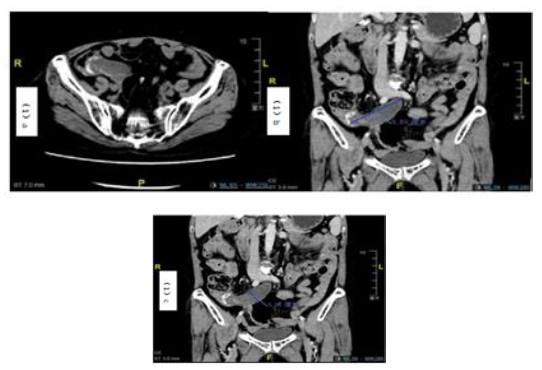
- enlarged appendix with intermittent enhancement points; b. appendix length close to 9 cm; c. appendix transverse diameter approximately 3 cm.
Figure 1: Preoperative abdominal enhanced computed tomography (CT) results of the patient
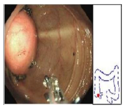
Figure 2: Colonoscopy results of a 3.5 cm × 3 cm mass at the ileocecal region
Laparoscopic appendectomy was performed after considering appendiceal mucinous adenocarcinoma. The appendix, located in the lower part of the cecum, was enlarged (approximately 6 cm × 3 cm × 3 cm), had a smooth surface but hard texture, and adhesion with the surrounding area was not obvious. After severing the appendiceal mesentery, the appendix was ligated and removed, and the specimen was full of mucus (Figure 3a, 3b, 3c, 3d).
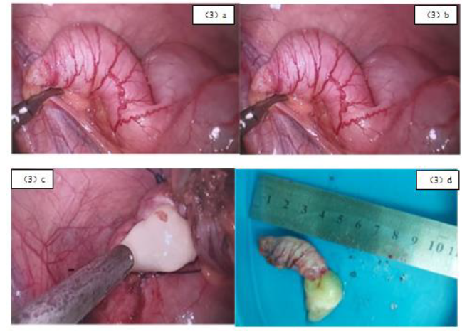
a, b: thickened appendix during operation; figure (3) c: yellow and white jelly after amputation of appendix during operation; figure (3) d: the specimen of appendix was removed after operation.
Figure 3: Appendix evaluation during surgery
A rapid frozen section examination was done and the results suggested appendiceal mucinous tumor. Additionally, a laparoscopic right hemicolectomy was performed, dissociating upward to the horizontal plane of the duodenal bulb via the caudal approach, outward to the right paracolic sulcus, upstream to the hepatic flexure of the colon, fully dissociating the cecum and terminal ileum (15 cm), and resecting the ascending colon. Ileocolonic end-to-side anastomosis was performed to close the abdominal cavity, and then the patient received cisplatin together with intraperitoneal hyperthermic perfusion.
Postoperative pathological examinations of the appendix were performed. The results showed a dilated lumen, villous or cluster hyperplasia of the local lining epithelium, mild to moderate atypia associated with epithelial cells, enlarged and deeply stained focal epithelial nuclei, easily observed mitotic images, and the disappearance of intrinsic glands. Moreover, the muscular layer of the mucosa was not obvious, and the wall thickened with fibrous tissue hyperplasia. The formation of mucus paste in the lumen (between the smooth muscle of the vessel wall and the serous fibrous fat), and some free mucus tissue were observed. In appendiceal mucinous tumors, most of the epithelium show low-grade changes, whereas focal epithelial changes show high-grade alterations; they tend to progress to high-grade mucinous adenocarcinomas of the appendix, and the tumor tissues encompass the whole layer of the appendix to the outer serosa. Part of the free mucus tissue was also seen (Figure 4a). The root of the appendix showed lumen-like tissue, smooth muscle, no mucosal epithelium, local calcification, and no clear tumor tissue (Figure 4b).
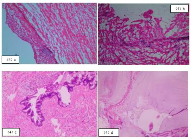
Figure 4a, b: pathological results of rapid freezing of appendix tissue during operation; figure (4) c: postoperative pathological results of appendix tissue; figure (4) d: pathological results of ileocecal intestine after operation.
Mucinous tumor components were not found in the mucous layer and muscular layer of the intestinal wall. Mucus and calcified tissue were seen in the serous layer of the local intestinal wall, which was considered to be involved in appendiceal mucinous tumors; tumor tissues were not found in the lateral incisal margin of the small intestine and large intestine. Four lymph nodes were examined but no tumor tissues were found (Figure 3C). Immunohistochemical results showed positivity for cytokeratins (CK), CD X Mel 2, carcinoembryonic antigen (CEA), Ki-67 protein (approximately 20% +), and a few p53 cells. On the fifth day after the surgery, the abdominal drainage fluid suddenly increased, the color intensity increased to bright red, but there were dark red exudates at the orifice of the drainage tube, and flaky dark red subcutaneous ecchymosis could be seen in the waist and abdomen. Check Fibrinogen(Fbg )0.97 g/L, and fibrinogen was infused with 10 units of cryoprecipitate, and Fbg 0.80 g/L, 0.92 g/L, and PT 14.7 seconds. PT/R 1.29, PT international normalized ratio 1.30, PT% 57.2%, TT 21.3 seconds, D-Di 15.99 (mg/L) (Figure 5a, 5b, 5c, 5d, 5e, 5f, 5g for specific changes and distribution between groups before and after surgery).
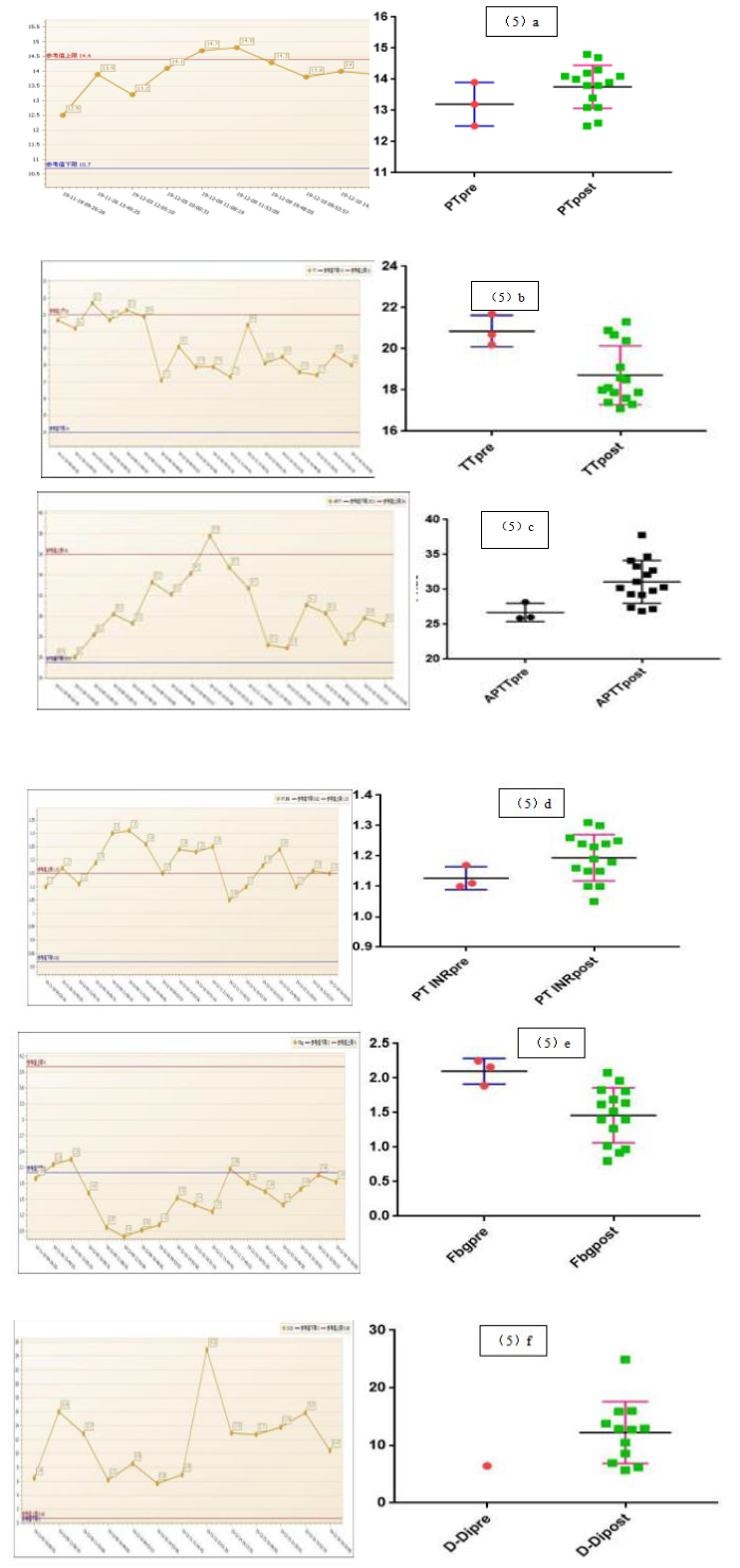
Figure 5a: patient PT trend chart and preoperative and postoperative PT group comparison; Figure 5b: patient APTT trend chart and preoperative and postoperative APTT group comparison; Figure 5c: patient TT trend chart and preoperative and postoperative TT group comparison; Figure 5d: patient PT INR trend chart and preoperative PT INR comparison between groups; Figure 5e: patient Fbg trend chart and preoperative Fbg comparison between groups; Figure 5f: D-Di trend chart of patients and comparison between D-Di groups before and after operation; Figure 5g: PT, TT, APTT, Fbg, PT INR, D-Di of patients.
The drainage fluid from the abdominal cavity and pelvic drainage tube gradually increased, and the color deepened. Concurrently, the hemoglobin concentration decreased gradually from 115 g/L to 90 g/L, 71 g/L, and 64 g/L after the surgery. Blood concentration was considered, but the color and volume of drainage fluid changed, and the concentration of hemoglobin decreased greatly. Chronic abdominal bleeding was investigated, and disseminated intravascular coagulation could not be ruled out. Cryoprecipitate, virus-inactivated plasma, suspended red blood cells, and human serum albumin injection were infused. Simultaneously, symptomatic support treatment such as aminomethylbenzoic acid hemostasis and intravenous injection of vitamin K to improve blood coagulation function and strengthen anti-infection were administered, while fibrinogen injection was infused to maintain the blood fibrinogen level at approximately 1.50 g/L. After active conservative treatment, the abdominal and pelvic drainage of the patient decreased, and the color gradually faded. On the ninth day after the surgery, the concentrations of hemoglobin, D-Di, and Fbg were 88 g/L, 24.92 mg/L, and 1.81. g/L, respectively. During this period, 40 g of human albumin injection, 7 g of Fbg, 1.5 units of suspended red blood cells, 26 units of cryoprecipitate, and 300 mL of virus-inactivated plasma were infused. Nineteen days after the surgery, the patient was in a stable condition, and the abdominal cavity and pelvic drainage tube were removed; she recovered and was discharged from the hospital.
3. Discussion
The clinical manifestations of primary appendiceal tumors include lower abdominal distension and discomfort, muscle tension and rebound pain, and right lower abdominal mass. Due to the gradual enlargement of the tumors, the stricture of the root of the appendix or the extrusion of fecal stone leads to the obstruction of secretions in the appendiceal cavity. It causes an increase in intraluminal pressure and distension and discomfort of the lower abdomen [9-10]. Some tumors are larger than others and may invade the greater omentum, resulting in omentum-like masses. Some symptoms such as weight loss, intestinal obstruction, changes in defecation habits (acute weight and inexhaustible feeling), oliguria (compression of the right ureter leading to low urine output), anemia (hematochezia and chronic consumption of the body), distension of the lower abdomen in females, and ascites are misdiagnosed, thus delaying the timing of treatment [11]. The study [12] showed that the clinical misdiagnosis rate of primary appendiceal tumors is as high as 97.6%. This high misdiagnosis rate of primary appendiceal tumors may be due to (1) the low incidence of the disease (2) the inexperience of clinicians and a lack of morphological understanding of the rare disease; (3) the lack of diagnostic criteria due to atypical clinical symptoms of the disease. This patient was admitted to the hospital with cholecystolithiasis and cholecystitis without specific abdominal symptoms, abdominal examination without signs of peritonitis, and no tenderness or rebound pain at McDonnell's point. Contrast-enhanced CT indicated an appendiceal tumor and the disease was detected. Due to the lack of specific clinical manifestations of the disease, there is no definite conclusion on the diagnostic criteria. Serum CEA and CA19-9 are significant tumor markers that aid in diagnosis, but they are not specific [13]. In appendiceal mucinous gland tumors, the increase in CEA and CA19-9 was approximately 58% and 67%, respectively [14-15]. Alpha fetoprotein (329.6 ng/mL), CA19-9 (219.40 U/mL), and CEA (3.7 U/mL) were reported at the time of admission. B-mode ultrasonography can be used to screen suspected appendiceal mucinous tumors, and CT is often used as an important assistant tool for preoperative diagnosis. CT of appendiceal mucinous gonadal tumors is often characterized by an enlarged appendix, a circular or tubular cystic low-density shadow in the ileocecal part, thin and smooth cyst wall, soft tissue mass, and a clear surrounding mass. Some of the surrounding adipose tissues will show stripes and calcification, but rarely cecal invasion, cecal wall thickening, and exudation [16]. Clinically, these tumors are diagnosed by pathology. Surgical treatment is the first choice of treatment for the disease. During the surgery, the patient had an enlarged appendix (6 cm × 3 cm × 3 cm) located in the lower part of the cecum, with a smooth surface, hard texture, and no obvious adhesion with the surroundings. After double ligation of the appendix, the appendiceal mesentery was cut off and the appendix was removed. The specimen which was full of mucus was sent for rapid frozen pathological examination. The results were suggestive of an appendiceal mucinous tumor. After laparoscopic right hemicolectomy, the caudal approach was used to dissociate upward to the horizontal plane of the duodenal bulb, outward to the right paracolic sulcus, and upstream to the hepatic flexure of the colon, fully dissociating the cecum and the left and right ileum of the terminal 15 cm and cutting off the end of the ileum at the 12 cm of the ileocecum. After resection of the ascending colon, end-to-side ileocolonic anastomosis was performed. Hot perfusion irrigation tubes were placed in the left upper abdomen, right upper abdomen, left pelvic cavity, and right pelvic cavity, respectively. After preheating by the perfusion machine, the water temperature was constant at 42 °C, with both sides of the pelvic drainage tube as the inlets, and the left and right epigastric drainage tubes as the outlets. The patient was intraperitoneally infused with normal saline containing cisplatin solution for 60 min. Ito [17] studies have shown that the 5-year survival rate of unoperated patients with appendiceal mucinous gland tumors is about 32%, and the this rate is much higher in patients undergoing radical surgery than in patients who do not undergo surgery. With regard to the choice of surgical methods, Zeng [18] believes that all layers of the appendix are thin, and most of the tumors are located at the root of the appendix, and appendectomy alone is difficult to ensure complete tumor resection; therefore, it is suggested that once an appendiceal mucinous gland tumor is diagnosed, right colectomy is the next desirable procedure. If the tumor invades the whole layer and it is not clear whether there is peripheral lymph node metastasis, regional lymph node dissection should be performed. The scope of dissection can be referred to as D2 dissection of right colon cancer. If the surrounding omentum, peritoneum, and surrounding organs were invaded during the surgery, the invasion focus and organs were resected at the same time, and the mucus tissue was removed extensively and thoroughly. For female patients, it has been reported [16,19-21] that appendiceal mucinous tumors and ovarian mucinous adenocarcinomas can occur at the same time, and the two tumors are pathologically similar; therefore, it is not ruled out that ovarian tumors are the result of intraperitoneal implantation of appendiceal mucinous gland tumors. Hence, for female patients, the pelvic cavity must be explored during the surgery, and the ovaries must be evaluated to ascertain their condition, and quickly frozen for biopsy during the surgery, if necessary, to decide whether to have a one-stage resection. The surgery of patients with appendiceal mucinous tumors should be as slow as possible. Attention must be paid to the incision and surgical field, and intraoperative rupture of appendiceal tumor tissue resulting in cavity content flow out should be avoided, which might result in abdominal pseudomyxoid carcinoma [22]. During the surgery, the texture, size, activity, and adhesion of the tumor should be evaluated to avoid iatrogenic implantation caused by an improper surgery [15]. The temperature of hyperthermic intraperitoneal chemotherapy (HIPEC) perfusion solution can be maintained at 42 °C [23]. Most studies have confirmed that HIPEC can markedly improve the tumor-free survival time and total survival time of patients and reduce mortality [24-25]. Cisplatin, 5-FU, mitomycin, carboplatin, and other drugs are available for use as infusion drugs. There are data to prove that the 5-year survival rate of patients receiving HIPEC can be increased to approximately 97% [26]. For patients with extensive abdominal metastasis, systemic intravenous chemotherapy is feasible after surgery. At present, first-line drugs such as capecitabine, oxaliplatin, and irinotecan are available for colorectal cancer [10,27], but are ineffective in patients who are insensitive to the drugs; in such cases, the combination of radiotherapy and chemotherapy or targeted drug therapy can be considered if necessary [28]. Similar to other malignant tumors, appendiceal mucinous tumors can metastasize. Metastatic patterns are often infiltrative, and lymph node metastasis is visible, but blood metastasis is rare. In 2010, the World Health Organization classified primary appendiceal tumors as low-grade appendiceal mucinous tumors, mucinous adenomas (cystadenomas), low-grade peritoneal pseudomyxomas originating from the appendix, high-grade peritoneal pseudomyxomas originating from the appendix, appendiceal mucinous adenocarcinomas, and so on. The first three are non-invasive benign lesions, and the latter two are invasive malignant tumors. Among them, appendiceal mucinous adenocarcinoma is divided into three pathological types: (1) Polypoid type: the mass protrudes into the appendiceal cavity in the shape of a column, which can block the excretion of appendiceal contents to the cecum; (2) ulcerative type: local thickening and protuberance of the appendiceal wall, the formation of ulcerative surface, and bloody fluid exudation can be seen on part of the ulcerated surface; (3) infiltrative type: the local thickening of the appendiceal wall shows diffuse thickening, and tumor cells can be seen in the whole layer of its diffuse thickening. The patient had a pathological type of mucinous adenocarcinoma, with a high degree of malignancy and a poor prognosis; therefore, intraoperative HIPEC was performed during the surgery. Immunohistochemical analysis showed CK (+), CDX-2 (+), CEA (+), Ki-67 (approximately 20% +), and p53 (a few cells +). CEA is a group of acidic glycoproteins secreted by colorectal cancer cells, and it [29] is widely found in a variety of epithelial tumors (especially adenocarcinoma). It is generally believed that the lower the degree of tumor differentiation, the higher the positive expression rate of CEA [30], and the positive expression of CEA can be used as a predictor of lymph node metastasis [31-32]; the higher its expression, the greater the possibility of lymph node metastasis. A P53 mutant type lacks inhibition of cell proliferation and promotion of cell transformation and proliferation, thus, leading to tumor occurrence [30,33]. Concurrently, p53 can affect the prognosis of patients by affecting the sensitivity of tumor cells to chemotherapeutic drugs. The higher the level of p53, the worse the prognosis of patients [34]. As a sensitive nuclear antigen associated with cell proliferation, Ki-67 is abnormally expressed in a variety of tumors and precancerous lesions, which are closely related to the potential of tumor implantation, infiltration, and metastasis. Studies have shown that [35] Ki-67 can be used as an important index to evaluate the reproductive ability of cells, judge tumor proliferation, and distinguish benign from malignant tumors. The higher the content of Ki-67, the worse the prognosis. Therefore, the type of tumor determines patient prognosis. Carcinoids have the highest 5-year survival rate, followed by goblet cell carcinomas and mucinous adenocarcinomas. Signet ring cell carcinomas, highly malignant lesions in digestive tract tumors, have the lowest postoperative 5-year survival rate [36]. Therefore, combined with the results of pathological immunohistochemistry, it is suggested that patients should continue to receive adjuvant chemotherapy after surgery. In our case, the patient was plagued by a hepatitis B virus replication period. The virus causes inflammation and activates the immune response in the body, and regardless of whether the virus replicates or not, it will cause immune damage to the liver [37]. The postoperative abdominal bleeding was comprehensively considered as follows: (1) decreased immunity of the appendix as an immune organ; the patient underwent appendectomy, and had viral hepatitis, abnormal liver function, and loss of immune organs, resulting in low immunity. (2) the stress effect of anesthesia and surgery; surgery and anesthesia often aggravate the damage of platelet function, and the secondary enhancement of fibrinolytic activity in the perioperative period aggravates the original hyperfibrinolysis in patients with liver dysfunction [38]. Hypothermia induction of anesthesia and low hepatic blood perfusion during the surgery further reduced blood flow in the liver; surgical trauma caused the consumption of coagulation factors and platelets, resulting in excessive bleeding during and after the surgery. Moreover, liver dysfunction can lead to slow metabolism of narcotic drugs, which further aggravates the metabolic pressure of the liver and leads to a vicious circle in the process of blood coagulation in patients [39]. (3) the influence of coagulation factors V, VI, VII, IX, X, XI, XII, fibrinogen, and prothrombin all synthesized by the liver, and the abnormal liver function of the patients led to the disturbance of coagulation factor and fibrinogen synthesis, forming a vicious circle and further aggravating postoperative bleeding. (4) diffuse intravascular coagulation: tumor metastasis, a certain number of cancer cells entering the blood can activate coagulation factors through surface contact, thus activating the endogenous coagulation system and causing disseminated intravascular coagulation (DIC) [40], while DIC further aggravates the damage of liver function. (5) the patient was treated with cisplatin intraperitoneal thermal perfusion, the wound in the abdominal cavity had just formed a blood scab in the early stage after surgery, and the microvessels had not been fully mechanized. During HIPEC, a large amount of fluid was quickly injected into the abdominal cavity. Mechanical lavage and accelerated blood flow rate may affect the elastic contractility of blood vessels, resulting in chronic blood oozing from small blood vessels. Simultaneously, cisplatin perfusion may affect liver and kidney function, resulting in blood coagulation dysfunction. It is easy to cause intra-abdominal bleeding, while fibrinogen levels continued to decrease and D-dimer levels increased. Considering the production of D-Di after fibrinogen hydrolysis, the level of D-Di in blood increased, which further affected the blood coagulation function of patients. For the report of this disease, due to the small number of cases and lack of representation, the best treatment for the disease cannot be defined, and it still requires a large number of cases or multicenter clinical verification. It is suggested that the patients should be treated with adjuvant chemotherapy after surgery, but the patients cannot fully cooperate with the treatment and lack complete systematic treatment. To date, patients have a short survival time after treatment, and the prognosis has not been evaluated sufficiently; therefore, it still needs a long follow-up period.
References
- Zhuang ZH, Tan SC, Chen G, et al. Diagnosis and treatment of 3 cases of Adennocarcinoma of the Vermiform appendix. Chinese Journal of General Surgery 23 (2014): 530-532.
- Tran S, HolIoway RN. Metastatic appendiceal mucinous adenocarcinoma to well-differentiated diffuse mesothelioma of the peritoneal cavity: a mimicker of florid mesothe1ial hyperplasia in association with neoplasms. Int J Gynecol Pathol 27 (2008): 526-530.
- Harfi S, Sellami A, Affes A, et a1. Histopath0109ical findings in appendectomy specimens: A study of 24, 697 cases. Int J Colorectal Dis 29 (2014): 1009-1012.
- Conte CC, Petrelli NJ, Stulc J, et a1. Adenocarcinoma of the appendix. Surg Gynecol Obstet 166 (1988): 451-453.
- Ren XX, Xie FX, Tian Y, et a1. Clinical diagnosis and treatment of appendiceal mucinous adenocarcinoma. Moder Oncology 27 (2019): 0867 -0869 .
- Qi Zhaosheng, Liu Zhimin, Guo Wei. Practice of abdominal surgery. Beijing: China Medical Science and Technology Press (1996).
- Xu XL, Yang JH, Chao Y. Clinical and pathological chaeacteristics of gastrointestinal tumors related to schistosomiasis japonica. Tropical Diseases and Parasitology 12 (2014): 7-9.
- Heung K, Lee SS, Raman M. Prevalence and mechanisms of malnutrition in patients with advanced liver disease, and nutrition management strategies. Clin Gastroenterol Hepatol 10 (2012): 117-125.
- Cheng Y, Chen YX, Qin SK, et al. Linical analysis of 13 patients with primary appendix mucinous adenocarcinoma [J]. China Journal of Modern Medicine 20 (2010): 3370-3372.
- Fu J, Wang B, Jiang XF, et al. Diagnosis and surgical treatment of appendiceal mucinous tumor. Chin J Surg Onco 10 (2018): 190-192.
- Ma Y, Tu SP. Clinical Features and Survival Analysis of Primary Appendiceal Mucinous Neoplasm [J]. Chin J Gastroenterol 21 (2016): 662-667.
- Huang XF, Tang Y. Diagnosis and treatment of 30 cases of Adennocarcinoma of the Vermiform appendix. Chinese Clinical Oncology 10 (2005): 425-427.
- Zhang CF, Sun C, Li SD, et al. Appendiceal Mucinous Adenocarcinoma with Peritoneal Metastasis: A Case Report and Review of Literature. Chin J Gastroenterol 22 (2017): 124-126.
- Carmignani CP, Hampton R, Sugarbaker CE, et al. Utility of CEA and CA19-9 tumor markers in diagnosis and prognostic assessment of muxinous epithelial cancers of the appendix. Surg Oncol 87 (2004): 162-166.
- O’Donnell ME, Carson J, Garstin WI. Surgical treatment of malignant carcinoid tumours of the appendix. Clin Pract 61 (2007): 431-437.
- Xuan ZQ, Gao L, Hua C. Diagnosis and treatment of 30 cases with appendiceal mucinous tumors. Abdominal Surgery 32 (2019): 284-286.
- Ito H, Osteen RT, Bleday R, et a1. Appendiceal adenocarcinoma: long-termoutcomes after surgical therapy. Dis Colon Rectum 47 (2004): 474-480.
- Zeng YJ, Liu L, Luo ZT, et al. Analysis of appendix adenocarcinoma treatment (1 case report attached). Lingnan Modern Clinical Surgery 11 (2011): 364-366.
- Tan FB, Liu HL, Pei HP, et al. Diagnosis and treatment of 30 cases of Adennocarcinoma of the Vermiform appendix. Chin J Gastroenterol Surgery 20 (2017): 340-341.
- Li Q, Zhao R. Diagnosis and treatment of 5 cases with appendiceal mucinous tumors. International Journal of Surgery 37 (2010): 786-787.
- Hu JX, Shi QX, Xu YT, et al. Diagnosis and treatment of appendiceal mucinous tumors. Zhejiang Medical Journal 37 (2005): 372-373.
- Austin F, Mavanur A, Sathaiah M, et al. Aggressive management of peritoneal carcinomatosis from mucinous apdieeal neoplasms. Ann Surg Oncol 19 (2012): 1386-1393.
- Elias D, Honore C, Ciuchendea R, et a1. Peritoneal pseudomyxoma: results of a systematic policy of complete cytoreductive surgery and hyperthermic intraperitoneal chemotherapy. Br J Surg 95 (2008): 1164-1171.
- Kell YK J. Management of appendix cancer. Clin Colon Rectal Surgery 28 (2015): 247-255.
- Dube P, Sideris L, Law C, et al. Guidelines on the use of cytoreductive surgery and hyperthermic intraperitoneal chemotherapy in patients with peritoneal surface malignancy arising from colorectal or appendiceal neoplasms. Curr Oncol 22 (2015): e100-e112.
- Shaib wL, Martin LK, Choi MA, et a1. Hyperthermic intraperitoneal chemotherapy following cytoreductivesurgery improvesoutcome in patients with primary appenl mucinous adenocarcinoma: apooled analysis from ree tertiarycare centers. Oncologist 20 (2015): 907-914.
- Liu WL, Li DS. Clinical analysis 0f 17 patients with primary appendix mucinous adenocarcinoma. General Surgery and Clinical Journal of China 3 (2004): 122-123.
- Logan-Collins JM, Lowy AM, Robinson Smith TM, et al. VEGF expression predicts survival inpatiens with peritoneal surface metastases from mucinous adenocarcinoma of the appendix and colon. Annsurg Oncol 15 (2008): 738-744.
- Xu D, Li XF, JiangWZ. QIlantitaHVe realtime RT-PCR detection for CEA, CK20 and CKl9 mRNA in peripheral blood of colorectal cancer pahent. Zhejiang Univ Sci B 7 (2006): 445-451.
- GuGL, RenL, HuangRR, et al. ImmunohistocheIIIical expression of carcinoembryonic andgen, P53, nnl23, Ki-67, nndtidrug resistELnce. associated protein and their correlations with the clinicopathologv in colorectal carcinoma. Shiiie Huaren Xiaohua Zazhi 14 (2006): 2765-2770.
- Tocchi A, Costa G, Lepre L, et al. The role of serum and gastrric juice levels of carcinoembryonic antigen, CA19-9 and CA72-4 in patients with gastric cancer. Cancer Res Clin Oncol 124 (1998): 450-455.
- Ucar E, Semerci E, Ustun H, et al. Prognostic value of preoperative CEA, CA19-9, CA72-4, and AFP levels in gastric. Adv Ther 25 (2008): 1075-1084.
- Zhao DP, Ding XW, Peng P, et al. Prognostic significance of bcl-2 and P53 expression incolorectal carcinoma. Zhejiang Univ Sci B 6 (2005): 1163-1169.
- Ralhan R, Swain RK, Agarwal S, et al. P-glycoprotein is positively correlated with P53 in human oral pre-malignant and malignant leaion and is associated with poor prognosis. Int J Cancer 84 (1994): 80.
- Salminen E, Palmu S, Vahlberg T, et al. Increased proliferation activity measured by immunoreactive Ki-67 is associated wim sivival improvernent in rectal/recto sigmoid cancer, World J Gastroenterol 11 (2005): 3245-3249.
- Turaga KK, Pappas SG, Gamblin TC. Importance of hisogic subtype in the Staging of appendiceal tumors. Ann Surg Oncol 19 (2012): 1379-1385.
- Zhang B, Jin R, Chen EH. Histopathological analysis of livertissue of patients with normal liver function infected with hepatitis bvirus[J]. Clinical Focus 19 (2004): 24-26.
- Liu Hong, Xu Li, Gu Huaqun,et al. Clinical significance of perioperative measurements of fibrinolysis function in patients with liver diseases. Clin Anesthesiol 19 (2003): 395-396.
- Yao LN, Xue XH. Characteristic analysis of drug-induced liver diseases. Chin J Infect Dis 23 (2011): 165-166.
- Wang Zhenyi, Li Jiazeng, Ruan Changgeng. Basic theory and clinic of thrombosis and hemostasis. Shanghai: Shanghai Science and Technology Press (1996): 546.


 Impact Factor: * 4.2
 Acceptance Rate: 72.62%
  Time to first decision: 10.4 days
  Time from article received to acceptance: 2-3 weeks