The Effect of Missense Mutation of FAM20A on Dental Development and Mineralization
Itai Zilberman1, Uri Zilberman2*
1Senior Resident, Department of Maxillofacial Surgery, Barzilai Medical University Center, Ben-Gurion University of the Negev, Beer –Sheva, Israel
2Head of Pediatric Dental Clinic, Barzilai Medical University Center, Ben-Gurion University of the Negev, Beer –Sheva, Israel
*Corresponding Author: Dr. Uri Zilberman, Head of the Pediatric Dental Clinic, Barzilai Medical University Center, 2nd Hahistadrut st, 78278, Ashkelon, Israel
Received: 28 December 2020; Accepted: 19 January 2021; Published: 29 January 2021
Article Information
Citation:
Itai Zilberman, Uri Zilberman. The Effect of Missense Mutation of FAM20A on Dental Development and Mineralization. Archives of Clinical and Medical Case Reports 5 (2021): 193-203.
View / Download Pdf Share at FacebookAbstract
FAM20A is a Protein Coding gene. It acts as an allosteric activator of the Golgi serine/threonine protein kinase FAM20C and is involved in biomineralization of teeth. A novel homozygous deletion mutation in exon 11 of FAM20A has been described in five members from a large consanguineous Bedouin family, all five with hypoplastic-hypocalcified AI and unerupted and crown resorbed permanent molars, a very rare phenotype. Deciduous mandibular canines and first molar, and maxillary first and second molars of an affected girl were compared to normal pair-matched teeth. Slices of the teeth were examined using SEM, and the ion content of enamel and dentine was determined using Energy Dispersive X-ray Spectrometer (EDS). The deciduous teeth showed missing enamel and the dentin was hypercalcified with very few open tubuli. Calcium and phosphorous percentages were reduced by 45 % in the enamel, and extensive apposition of secondary dentin and calcification of the pulps was observed. The permanent dentition showed impacted molars, premolars and canines with hypoplastic enamel and reduced dimensions of the erupted teeth. The mutation of FAM20A caused a peculiar type of AI with very thin amorphic enamel, infraoccluded deciduous teeth and impacted permanent canines, premolars and molars.
Keywords
<p>FAM20A; Aprismatic Enamel; Dentin; Hypoplastic Amelogenesis Imperfecta; Pulp calcification; Impaction of permanent teeth</p>
Article Details
1. Introduction
Amelogenesis Imperfecta (AI)(MIM#301200) represents a broad spectrum of congenital disorders which present with a rare abnormal formation of enamel in both primary and permanent dentition. AI is due to the malfunction of the proteins in the enamel as a result of abnormal formation via amelogenesis [1-3]. It is the oldest hereditary disorder affecting enamel, observed in early hominins. It has been described in a Homo erectus child from Melka Kunture, Ethiopia (Garba IV) dated to circa 1.5 MY [4] and possibly as pitted AI in Paranthropus [5]. The defects in AI are classified according to their clinical appearances: Type 1: hypoplastic, Type 2: hypomaturation, Type 3: hypocalcified and Type4: hypomature-hypoplastic enamel with taurodontism [6]. The mildest problems occur in the pitted hypoplastic type whereas the most severe are encountered in the hypocalcified type of AI [7]. Hypoplastic AI is characterized by thin but fully mineralized enamel or even complete absence of enamel that results from failure during the secretory stage. Hypomaturation AI is caused by incomplete removal of protein from the enamel matrix and results in brittle enamel. Hypocalcified AI is characterized by insufficient transport of minerals into the developing enamel and results in soft enamel. The mean enamel mineral content in hypomature and hypocalcified AI is reduced, while in the hypoplastic variant, enamel mineral content is normal [8]. The hypocalcified AI may be caused in part by malfunction of matrix protein degradation genes (MMP20 and KLK4) during the apposition and maturation phases [9].
The FAM20 family contains three separate subfamilies which are referred to as FAM20A (NM_017565.3), FAM20B (NM_014864.3) and FAM20C (NM_020223.3) in humans [10]. FAM20A (FAM20A Golgi Associated Secretory Pathway Pseudokinase) is a Protein Coding gene. It acts as an allosteric activator of the Golgi serine/threonine protein kinase FAM20C and is involved in biomineralization of teeth. It forms a complex with FAM20C and increase the ability of FAM20C to phosphorilate the proteins that form the matrix that guide the deposition of the enamel minerals. FAM20A is expressed in secretory ameloblasts, maturation stage ameloblasts, suprabasal cells of the gingivae, odontoblasts, and dental pulp cells, indicating that FAM20A plays a fundamental role in enamel development and gingival homeostasis. Ameloblasts express FAM20A during the secretory stage, consistent with hypoplastic AI phenotype (type AI1g) [11]. All FAM20A mutations causing AI described to date are found in autosomal recessive forms. Most mutations lead to transcript degradation as a result of premature stop codons or alteration of splice sites [12, 13]. Autosomal recessive missense mutations in FAM20A have been found in association with gingival fibromatosis and as part of the enamel renal syndrome [11]. Both syndromes are phenotypic variants of the same condition and are termed ERS (OMIM#204690) [14]. A novel homozygous deletion mutation in exon 11 of FAM20A (an A>T substitution followed by a single base deletion) has been described in five members from a large consanguineous Bedouin family, all five with hypoplastic-hypocalcified AI and unerupted and crown resorbed permanent molars, a very rare phenotype [15]. We compared the ultrastructure and mineral content of deciduous teeth from the girl with autosomal recessive hypoplastic AI due to missense mutation of FAM20A to match-paired normal deciduous teeth.
2. Patient Data
Sixteen years-old girl (AS) from the Bedouin family, with the novel homozygous deletion mutation in exon 11 of FAM20A, was examined and treated at a pediatric dental clinic, with a major complaint of "ugly teeth". Apart from the dentition pathology, her medical history and physical examination were normal. Intra oral examination revealed erupted permanent teeth with reduced mesio-distal and bucco-lingual dimensions, with no observed enamel and with yellow discoloration (Figure 1a-b). The panoramic view (Figure 1c) showed very thin enamel on the unerupted second molars and premolars, missing enamel on the deciduous teeth, infraoccluded deciduous primary molars and impacted ankylosed permanent canines, premolars and second molars. The pulp of the deciduous teeth showed massive calcification. The final diagnosis was hypocalcified-hypoplastic Amelogenesis Imperfecta with unerupted permanent teeth and crown resorption. The final treatment included extraction of all deciduous teeth, filling the gaps with artificial bone and restoration of occlusion with ceramic fused to metal crowns (Figure 1d).
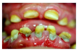
Figure 1a: Clinical appearance of the teeth of the girl with FAM20A mutation, front view.
Note the yellowish color due to hypoplastic enamel and the reduced dimensions of the teeth.
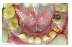
Figure 1b: Clinical appearance of the teeth of the girl with FAM20A mutation, lower jaw.
Note the very thin enamel, the infraoccluded first deciduous molars and the unerupted ankylosed permanent molars, premolars and canines.
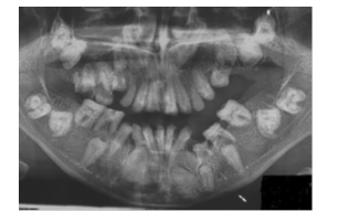
Figure 1c: Diagnostic dental panoramic views of the girl with FAM20 mutation.
The deciduous teeth were extracted, artificial bone substrate was inserted in the locations and ceramic fused to metal bridges were cemented.
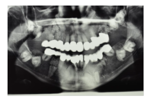
Figure 1d: Panoramic X-ray at the end of the treatment.
3. Teeth Preparation and Analyses
The teeth examined were the mandibular deciduous canine and first molar and maxillary deciduous first and second molars. The teeth were extracted during routine dental treatment and matched pair teeth from normal healthy children were collected after extractions during orthodontic treatment or after normal exfoliation. The parents gave their verbal consent to donate the teeth to the clinic. The teeth were invested in epoxy resin (Epofix Resin, Struers Co, Cleveland OH, USA) and sliced along a bucco-lingual plane parallel to the long axis of the tooth using an Isomet 1000 (Buehler Co, IL, USA). The deciduous canines were sectioned through the cups tip, and the deciduous first and second molars were sectioned through the mesial cusps. The sections were then examined using a scanning electron microscope (SEM, FEI, Quanta 200, Oregon, USA) under high vacuum mode, without coating.
Using Energy Dispersive X-ray Spectrometer (EDS), ion analyses were carried out for enamel and dentine. The
location of the ion analyses was identical in FAM20A and normal teeth. The relative ion concentrations were determined from a 150x150 microns rectangle with a minimum of 8000 counts.
3. Results
A very thin amorphous enamel layer was detected on the deciduous mandibular affected canine on the buccal surface close to the CEJ (Cemento-Enamel Junction) (Figure 2a) in comparison with a deciduous canine from a healthy girl (Figure 2b). No enamel layer was observed on the affected deciduous molars. A first deciduous affected molar (Figure 3a) was compared to a normal tooth (Figure 3b). The crown of the affected deciduous canine and molar showed an extensive secondary dentine formation and a distinctive breakdown line between the primary (D1) and secondary dentin (D2). On the deciduous canines full obliteration of the pulp canal occurred (D3) and in the molars a small residual pulp chamber could be observed. SEM images of enamel and dentin of affected canine (Figure 4a) and first deciduous molar (Figure 5a) is shown in comparison with match-paired normal teeth (Figure 4b, 5b).
Examination of the SEM images at high magnification showed major differences in both enamel and dentine development between the normal and affected deciduous teeth. In the normal enamel (Figure 4b) the prisms are arranged regularly, with large inter-prismatic layer between them, while in the deciduous canine from the affected girl the enamel is aprismatic and amorphous (Figure 4a). In normal dentine the tubuli form a regular orderly pattern (Figure 5b), in comparison with very few open tubuli and hypercalcified dentine in the affected canine (Figure 5a), and pulp calcifications (Figure 5c). Similar results were observed for the affected second deciduous molar.
Table 1 summarizes the main elements in enamel and dentine in the deciduous canines and deciduous first and second molars. In the enamel of the deciduous canine, the relative calcium and phosphate content was reduced by 54-55% relative to the normal tooth. The relative content of carbon was increased by 450% and of oxygen by 40%. In the deciduous canine dentin the relative content of carbon was increased and the content of calcium was reduced. The nitrogen content in D1 and D2 was increased in comparison with normal dentin. No significant differences were found between the primary (D1) and secondary (D2) dentin content. In the pulp calcification (D3) a reduced content of carbon and nitrogen was observed in comparison with primary or secondary dentin. In the deciduous molars the relative content of carbon and oxygen in both primary and secondary dentin are lower in comparison with normal dentin and the relative content of phosphate and calcium were higher.
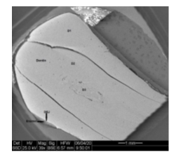
Figure 2a: SEM figure of the slice of affected mandibular deciduous canine.
Note: Very thin enamel layer only on the lingual aspect of the canine, D1- primary dentin, D2-secondary dentin, D3- pulp calcification.
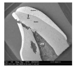
Figure 2b: SEM figure of the slice of normal deciduous canine.
Note total missing of enamel, the massive apposition of secondary dentin and calcifications of the pulp chamber.
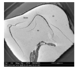
Figure 3a: SEM figure of first deciduous molar the girl affected by mutation of FAM 20A.
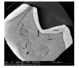
Figure 3b: SEM figure of normal first deciduous molar.
Note: Amorphous and aprismatic enamel.
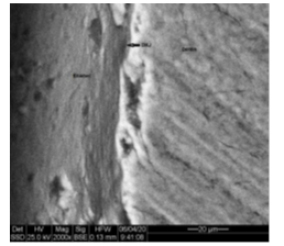
Figure 4a: Enamel of FAM20 deciduous canine.
Note: Prisms and inter-prismatic layer.
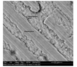
Figure 4b: SEM pictures of normal enamel of deciduous canine X2000.
Note: D1-primary dentin, D2-secondary dentin, and very few tubuli openings.
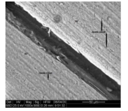
Figure 5a: Dentin of FAM20 deciduous canine, X1000.
Note: normal dentin tubuli.
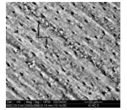
Figure 5b: SEM pictures of normal dentin of deciduous canine, X2000.
Note: Total obliteration of the pulp chamber and no dentin tubuli.
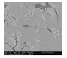
Figure 5c: Pulp (D3) area of primary canine of FAM20A girl.
|
Nitr |
Nitr |
Calc |
Calc |
Phos |
Phos |
Oxy |
Oxy |
Carb |
Carb |
|
|
|
FAM |
N |
FAM |
N |
FAM |
N |
FAM |
N |
FAM |
N |
|
Tooth |
|
0.00 2.03 1.98 1.31 |
0.00 1.45 |
30.41 49.54 47.06 48.35 |
55.59 52.71 |
11.24 20.22 20.61 19.15 |
20.70 19.69 |
21,27 17.79 20.39 20.15 |
15.49 18.74 |
37.08 10.31 10.18 9.51 |
8.22 7.42 |
E D1 D2 P |
Lc |
|
2.23 1.08 3.34 |
0.00 3.57 |
45.41 47.86 33.62 |
42.79 28.85 |
19.59 19.94 13.46 |
20.44 16.30 |
22.22 20.86 19.21 |
26.41 32.51 |
10.55 11.34 30.37 |
10.33 18.77 |
E D1 D2 P |
Ld |
|
2.02 1.03 |
0.00 1.92 |
43.25 52.90 |
40.22 27.08 |
20.20 19.19 |
20.69 16.78 |
22.55 19.73 |
29.81 37.91 |
11.95 10.20 |
7.42 16.32 |
E D1 D2 |
Ud |
|
1.98 0.00 |
0.00 2.36 |
52.90 54.12 |
52.45 45.34 |
20.29 20.78 |
20.97 19.14 |
16.15 16.53 |
19.93 22.73 |
8.67 8.57 |
6.65 10.43 |
E D1 D2 |
Ue |
Note: Lc= Mandibular deciduous canine, Ld= Mandibular deciduous first molar, Ud= Maxillary deciduous first molar, Ue= Maxillary deciduous second molar, Carb=Carbon, Oxy=Oxygen, Phos=Phosphate, Calc=Calcium, Nitr=Nitrogen, N=Normal, FAM=Fam20A mutation, E=Enamel, D1= Primary Dentin, D2=Secondary dentin, P=Pulp calcification.
Table 1: Relative ion concentration (Wt%) in deciduous teeth of FAM20A mutation compared to normal teeth.
4. Discussion
A detailed review of the literature regarding the correlation between the clinical phenotype of AI and enamel thickness and ultrastructure was published by Zhang and colleagues [16]. In localized hypoplastic AI the enamel was of normal thickness with disrupted prism morphology. In generalized hypoplastic AI the enamel was thin and lacked a prismatic structure. In hypomaturation AI the enamel was of normal thickness in males and thin in females and lacked a prismatic structure with mineralized deposits. In hypocalcified AI, the enamel thickness is normal or reduced and lacks the typical prismatic structure with irregular, poorly formed rods and with areas of amorphous non-crystalline material [16]. In this case, the enamel was missing from most of the affected deciduous teeth examined. The dentine was amorphous with a large number of cracks and few open tubuli.
FAM20A is expressed in mice ameloblasts only during the secretory stage and in the odontoblasts. In FAM20A knockout mice, the enamel resembles human hypoplastic AI, leading to pitted, thin and disorganized enamel detached from the DEJ, but without apparent dentine defects [17, 18]. It differs from the case described here where the enamel was amorphous, very hypoplastic and hypomineralized in the deciduous canine and molars, and the dentine was hypermineralized. A large amount of secondary dentin was laid and a significant pulp calcification occurred. The dentin tubuli were occluded.
Energy-dispersive X-ray spectroscopy, (EDS), was used successfully to analyze differences in chemical characterization between healthy teeth and Down's or CP teeth and to analyze the effect of dental materials on chemical composition of enamel or dentine [19, 20]. The relative ion content in the enamel (in MW%) of the affected deciduous canine showed a significantly reduced calcium and phosphorus and increased carbon and oxygen content. The results differ significantly from the results described for a deciduous molar with AI in a FAM83H mutation where the calcium and phosphorus content was even lower [16]. The hypercalcified dentine described here, in FAM20A deciduous teeth is similar to the results observed in hypocalcified AI [21].
Acknowledgement
We want to thank Roxana Golan from the Isle Katz Institute for Nanoscale Science and Technology, Ben Gurion University of the Negev, Beer-Sheva for the work on the SEM and the EDS analysis of the ions content.
References
- Crawford PJ, Aldred M, Bloch-Zupan A. Amelogenesis Imperfecta. Orphanet J Rare Dis 2 (2007): 17.
- Kida M, Ariga T, Shirakawa T, et al. Autosomal-dominant hypoplastic form of amelogenesis imperfecta caused by an enamelin gene mutation at the exon-intron boundary. J Dent Res 81 (2002):738-742.
- Smith CE, Murillo G, Brookes SJ, et al. Deletion of amelotin exons 3-6 is associated with amelogenesis imperfecta. Human Mol Genet 25 (2016): 3578-3587.
- Zilberman U, Smith P, Piperno M, et al. Evidence of amelogenesis imperfecta in an Early African Homo erectus. J Hum Evol 46 (2004): 647-653.
- Towle I, Irish JD. A probable genetic origin for pitting enamel hypoplasia on the molars of Paranthropus robustus. J Hum Evol 129 (2019): 54-61.
- Aldred MJ, Savarirayan R, Craford PJ. Amelogenesis imperfecta: a classification and catalogue for the 21st century. Oral Dis 9 (2003): 19-23.
- Seow WK. Clinical diagnosis and management strategies of amelogenesis imperfecta variants. Pediatr Dent 15 (1993): 384-393.
- Wright JT, Hall K, Yamauche M. The enamel proteins in human amelogenesis imperfecta. Arch Oral Biol 42 (1997): 149-159.
- Takagi Y, Fujita H, Katano H, et al. Immunochemical and biochemical characteristics of enamel proteins in hypocalcified amelogenesis imperfecta. Oral Surg Oral Med Oral Pathol Oral Radiol Endod 85 (1998): 424-430.
- Nalbant D, Youn H, Nalbant SI, et al. FAM20: an evolutionarily conserved family of secreted proteins expressed in hematopoietic cells. BMC Genom 6 (2005): 11.
- Wang SK, Reid BM, Dugan SL, et al. FAM20A mutations associated with enamel renal syndrome. J Dent Res 93 (2014): 42-48.
- Cho SH, Seymen F, Lee KE, et al. Novel FAM20A mutations in hypoplastic amelogenesis imperfecta. Hum Mutat 33 (2012): 91-94.
- Kantaputra PN, Bongkochwilawan C, Kaewgahya M, et al. Enamel renal gingival syndrome, hypodontia and a novel FAM20A mutation. Am J Med Genet A 164A (2014): 2124-2128.
- Cui J, Xiao J, Tagliabracci VS, et al. A
- Nalbant D, Youn H, Nalbant SI, et al. FAM20: an evolutionarily conserved family of secreted proteins expressed in hematopoietic cells. BMC Genom 6 (2005): 11.
- Wang SK, Reid BM, Dugan SL, et al. FAM20A mutations associated with enamel renal syndrome. J Dent Res 93 (2014): 42-48.
- Cho SH, Seymen F, Lee KE, et al. Novel FAM20A mutations in hypoplastic amelogenesis imperfecta. Hum Mutat 33 (2012): 91-94.
- Kantaputra PN, Bongkochwilawan C, Kaewgahya M, et al. Enamel renal gingival syndrome, hypodontia and a novel FAM20A mutation. Am J Med Genet A 164A (2014): 2124-2128.
- Cui J, Xiao J, Tagliabracci VS, et al. A secretory kinase complex regulates extracellular protein phosphorylation. Elife 4 (2015): e06120.
- Volodarsky M, Zilberman U, Birk OS. Novel FAM20A mutation caused autosomal recessive amelogenesis imperfecta. Arch Oral Biol 60 (2015): 919-920.
- Zhang C, Song Y, Bian Z. Ultrastructural analysis of the teeth affected by amelogenesis imperfecta resulting from FAM83H mutation and review of the literature. Oral Surg Oral Med Oral Pathol Oral Radiol 119 (2015): e69-e76.
- Vogel P, Hansen GM, Read RW, et al. Amelogenesis imperfecta and other biomineralization defects in FAM20A and FAM20C null mice. Vet Pathol 49 (2012): 998-1017.
- Li L, Saiyin W, Zhang H, et al. FAM20A is essential for amelogenesis, but is dispensable for dentinogenesis. J Molecular Hystol 50 (2019): 581-591.
- Keinan D, Smith P, Zilberman U. Microstructure and chemical composition of primary teeth in children with Down syndrome and cerebral palsy. Arch Oral Biol 51 (2006)0: 836-843.
- Mass E, Hassan A, Zilberman U. Long term in vivo effects of various restorative materials on enamel and dentin of primary molars. Quintessence Int 48 (2017): 633-638.
- Sanchez-Quevedo MC, Ceballos G, Garcia JM, et al. Dentine structure and mineralization in hypocalcified amelogenesis imperfecta: a quantified X-ray histochemical study. Oral Dis 10 (2004): 94-98.


 Impact Factor: * 5.3
 Acceptance Rate: 75.63%
  Time to first decision: 10.4 days
  Time from article received to acceptance: 2-3 weeks
