Three Critical COVID-19 Cases Treated with Mesenchymal Stem Cells: A Case Series Report
Karakoc ZC1, Koyuncu Irmak D2, 3*, Tugrul S4, Ece F5, Gormeli CA6, Ozyilmaz K4, Karaoz E2, 3, 7, 8
1Department of Infectious Diseases and Clinical Microbiology, Istinye University, Istanbul, Turkey
2Department of Histology & Embryology, Istinye University, Istanbul, Turkey
3Stem Cell and Tissue Engineering R&D Center, Istinye University, Istanbul, Turkey
4Department of Anaesthesia and Intensive Care, Istinye University, Istanbul, Turkey
5Department of Chest Diseases, Istinye University, Istanbul, Turkey
6Department of Radiology, Istinye University, Istanbul, Turkey
73D Bioprinting Design and Prototyping R&D Center, Istinye University, Istanbul, Turkey
8Center for Stem Cell and Regenerative Therapies (LivMedCell), Liv Hospital/Ulus, Istanbul, Turkey
*Corresponding Author: Duygu Koyuncu Irmak, Department of Histology and Embryology, Istinye University, School of Medicine, Istanbul, Turkey
Received: 29 October 2020; Accepted: 07 December 2020; Published: 04 January 2021
Article Information
Citation: Karakoc ZC, Koyuncu Irmak D, Tugrul S, Ece F, Gormeli CA, Ozyilmaz K, Karaoz E. Three Critical COVID-19 Cases Treated with Mesenchymal Stem Cells: A Case Series Report. Archives of Clinical and Medical Case Reports 5 (2021): 008-042.
View / Download Pdf Share at FacebookAbstract
This paper reports three critically ill patients diagnosed COVID-19 infection who were treated with conventional therapy and MSCs add-on therapy in combined treatment protocols on a patient centric manner. We followed up and evaluated the cases specifically for efficacy and safety indicators. We introduce promising efficacy and safety data of the add on MSC therapy in COVID-19. We believe that the MSC therapy could be a safe effective therapy method for novel coronavirus as add-on therapy and when applied to the critically ill COVID-19 cases on a personalized approach.
Keywords
<p>Covid-19; Novel coronavirus; Cell-based therapy; Combined therapy</p>
Article Details
Abbreviations:
SARS-CoV- Severe acute respiratory syndrome coronavirus; ARDS- Acute Respiratory Distress
Syndrome; MOD- Multiorgan dysfunction; MSCs- Mesenchymal stem cells; RT-PCR- Real Time Polymerase Chain reaction; CT- Computed tomography; BUN- Blood urea nitrogen; WHO- World Health Organization; CPls- Convalescent Plasma; PT- prothrombin time; aPTT- activated partial thromboplastin time; AST- Aspartate transaminase; ALT- Alanine transaminase; CRP- C-reactive protein; PCT- Procalcitonin; CP- Convalescent Plasma
cGMP- Current Good Manufacturing Practices; ICU- Intensive Care Unit;MRSE- Methicillin resistant staphylococcus epidermis; MRSH- Methicillin resistant staphylococcus hominis
1. Introduction
A new pathogen, novel coronavirus (SARS-CoV-2) burst in China on Dec 2019 and spread over the world only in one month [1]. Since that time, the world is learning new living norms of being protected from the new disaster which has been still a threat for the public health [2, 3]. There is no cure or vaccine for this disease yet, however, there have been huge effort in investigating the therapeutic approaches to reduce the inflammatory response, to prevent or treat the secondary bacterial and/or viral infections with corticosteroids, antibiotics, antivirals, convalescent plasma, even some anti-parasites, and anti-malarial medicines, besides aggressive standard supportive care [4-7]. The cell-based therapies, particularly involving MSCs, have been attracting growing interest for treating COVID-19 caused respiratory disease, resulting several clinical trials of varying clinical development phases registered from a lot of locations of the world (https://www.who.int/ictrp/search/en/). Despite being limited, there are some clinical trial results published in the medical literature revealing the promising curing outcomes [8, 9]. Besides, there have been several more case reports published in which the COVID-19 diagnosed cases treated with MSCs, also reporting this regenerative advanced medicinal product demonstrate certain efficacy in treating this disease, especially in critically ill stage patients [10-12]. MSCs have been investigated in many medical conditions and ARDS experimentally and clinically [13]. With the immunomodulatory and anti-inflammatory properties, as well as angiogenic and even reconstructive characteristics of MSCs make them appealing in consideration of potential promising cure for ARDS treatment [14-19]. In this paper, we report three critically ill patients diagnosed COVID-19 infection admitted to the Istinye University affiliated Liv Hospital 1-18 April 2020 who were treated with MSCs as add-on therapy in combined treatment protocols. We followed up and evaluated the cases specifically for efficacy and safety indicators and hereby we report all patients’ outcome in evidence-based manner.
2. Materials and Methods
2.1 Treatment decisions
Combined treatment protocol decisions for the patients were made based on the international literature and the guideline published and regularly updated by the Scientific Advisory Committee which has been assembled in the Ministry of Health in Turkey, specifically for the COVID-19 management purposes right after the WHO declaration of ‘pandemic’ [20, 21]. Upon admission to the hospital, all three patients were immediately checked serologically and confirmed as Hepatitis B, C, Influenza A/B virus, and Human Immune-Deficiency Virus negative. The treatment consisting of antibiotics, antivirals, corticosteroids, supporting and prophylactic treatments (such as anti-thrombolytics, antipyretics, analgesics, proton-pump inhibitors, fluid replacement, etc.) was applied. Additionally, all patients received MSC treatment as add-on therapy upon ethical, scientific and regulatory approval of Turkish Health Authority. All the patients were consented via the legally authorized representatives by the treating physicians in compliance with the applicable regulations [22, 23]. For the overall treatment and MSC-add on therapy, the efficacy and safety indicators below were defined, collected and evaluated.
2.2 Primary efficacy indicators
- 1 Level of oxygenation
- 2 Indicators of thorax computed tomography (CT) images
- 3 RT-PCR results
2.3 Primary safety indicator
It was defined as local or systemic adverse events that are recorded throughout the hospitalization as clinically significant and MSC treatment related.
2.4 Secondary efficacy indicators (from biological samples)
- 1 White blood cells (WBC), lymphocytes, red blood cells (RBC) and platelets levels in hemogram of the patients
- 2 Liver function tests of AST, ALT, direct and total bilirubin levels
- 3 Kidney function tests of BUN, creatinine levels
- 4 Ferritin, Procalcitonin, CRP levels as inflammatory biomarkers
- 5 Coagulation factors (D- dimer, prothrombin time, activated partial thromboplastin time)
- 6 Blood, urine, tracheal aspirate culture results
2.5 Stem cell processing, quality control and preparation
All samples of Wharton Jelly derived mesenchymal stem cells (WJ-MSCs) as cell therapy medicinal products were isolated, expanded, analysed, and prepared in the cGMP-certified facility at Liv Hospital, Center for Regenerative Medicine and Stem Cell Manufacturing (LivMedCell) as described previously (24, 25).The MSCs were slowly drawn into the syringe without pressure having 40 ml volume and suspended in 150 ml of 0.9 % NaCl. The total volume of 190 ml reconstituted MSC treatment was applied at each of the treatment by intravenous infusion in 1 hour.
2.6 Laboratory and imaging evaluations
The following laboratory and imaging assessments were performed during patient follow up: level of oxygenation, thorax CT, RT-PCR results, local or systemic clinically significant adverse events that are recorded throughout the hospitalization, white blood cells (WBC), red blood cells (RBC) and platelets levels in hemogram of the patients, liver function tests of AST, ALT, direct and total bilirubin levels, kidney function tests of BUN, creatinine, ferritin, procalcitonin, CRP levels as inflammatory biomarkers, coagulation parameters of D- dimer, prothrombin time, activated partial thromboplastin time, and blood, urine, tracheal aspirate culture results for evaluation of secondary infections.
3. Results
3.1 Cases’ presentation
There are three cases included in this report who were diagnosed critically ill COVID-19 disease. All of them had confirmed COVID-19 respiratory disease, and has negative serology for the Hepatitis B, Hepatitis C, Influenza A/B virus and Human Immunodeficiency Virus infections.
3.1.1 Case 1: 55 years old male patient admitted to the hospital with complaints of cough, fatigue, high fever and respiratory distress on April 2, 2020 and hospitalized with the diagnosis of COVID-19 pneumonia. At the time of admission, the patient reported that he had been receiving hydroxychloroquine, oseltamivir and azithromycin for 4 days. The patient was a heavy smoker. As the signs compatible with bilateral pneumonia in thorax CT observed, the patient was transferred to the ICU and the treatment was started on the same day. The combined treatment regime with add-on MSC therapy applied for this patient, and the biological samples’ culture results along with RT-PCR results are demonstrated in the Figures 1 and 2, respectively. As the symptoms, laboratory and imaging indicators demonstrated the improvement, the patient was extubated on April 17, 2020 and started spontaneous respiration. The patient was taken to the in-patient clinics from ICU on 21 April 2020. The patient was discharged from the hospital on 7 May 2020, with recovery.
3.1.2 Case 2: 87 years old female patient admitted to the hospital at April 8, 2020 with cough, fever and shortness of breath complaints for five days to admission as evaluated along with the thorax CT results; the patient has been hospitalized at the same day. Patient had the co-morbidities of hypertension, cardiac arrythmia and use of cardiac pacemaker. At 14 April 2020, with immediately started treatments, the patient was transferred to ICU, and upon worsening, at 15 April 2020, the patient was intubated. The combined treatment regime with add-on MSC therapy applied for this patient, and the biological samples’ culture results along with RT-PCR results are demonstrated in the Figures 1 and 2, respectively. With the improved thorax CT and oxygen saturation with concurrent alleviated symptoms, the patient was extubated on 19 April 2020 as patient could perform spontaneous respiration and transferred to the in-patient clinics from ICU on 20 April. The patient was discharged with recovery on May 15, 2020.
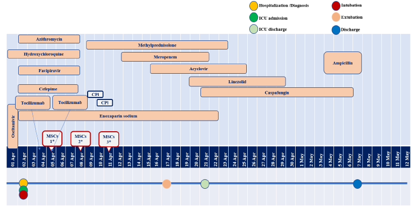
Figure 1: Case 1 Hospitalization and treatment schedules. (*: MSCs 1, 2, 3: MSC treatment Day 1, Day 2 and Day 3, respectively, at 270X106190ml, at 180X106 at 180X106190ml, as intravenous infusion).
3.1.3 Case 3: The male patient of 54 years of age admitted to the hospital with complaints of severe cough, fever and shortness of breath for 3-4 days on 18 April 2020. After confirmation of COVID-19 diagnosis based on the thorax CT, patient was hospitalized immediately. Upon worsened respiratory distress, the patient was transferred to ICU on 22 April. Despite the antibiotics, antivirals, and supportive treatments, the patient clinical condition got worsened and he was intubated on 28 April. This patient received the MSC treatment at a later stage compared to the other patients evaluated, when the clinical condition of the patient has not improved. The combined treatment regime with add-on MSC therapy applied for this patient, and the biological samples’ culture results along with RT-PCR results are demonstrated in the Figures 5 and 6, respectively. Patient remained intubated until 27 May 2020, when the laboratory values, thorax CT and clinical evaluation demonstrated improvement. The patient has demonstrated the recovery from the respiratory symptoms, and it was supported by clinical status and the thorax CT findings. The patient stayed in the hospital for 3 weeks for physiotherapy and discharged on 22 Jul 2020 with recovery.
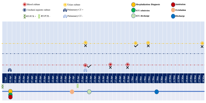
Figure 2: Case 1 Biological samples’ Cultures and RT-PCR assessments (20 April: Haemoculture showed Candida dubliniensis growth indicated the candidemia. 2 May: Urine culture showed Enterococcus faecalis growth was seen. These were evaluated as healthcare associated infections of candidemia and urinary tract infections).
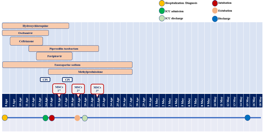
Figure 3: Case 2 Hospitalization and treatment schedules.(*: MSCs 1, 2, 3: MSC treatment Day 1, Day 2 and Day 3, respectively, at 120X106 190ml dose as intravenous infusion).
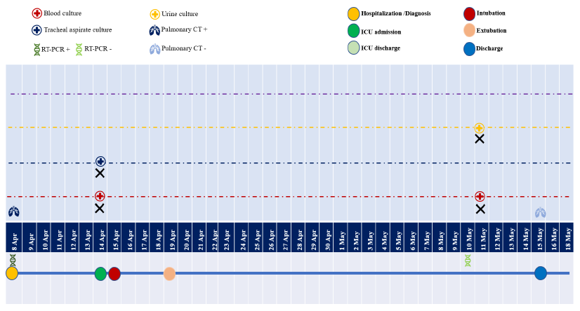
Figure 4: Case 2 Biological samples’ Cultures and RT-PCR assessments (there was no growth in any of the culture analyes.
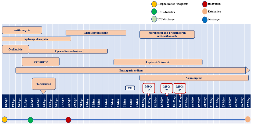
Figure 5: Case 3 KM Hospitalization and treatment schedules. .(*: MSCs 1, 2, 3: MSC treatment Day 1, Day 2 and Day 3, respectively, at 140X106 190ml dose as intravenous infusion).
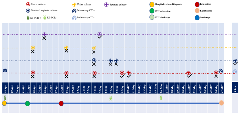
Figure 6: Case 3 Biological samples’ Cultures and RT-PCR assessments. (9 May; hemoculture, MRSE growth was seen as Catheter related bloodstream infection indicator. On 21 May hemoculture MRSH growth and on 9 June; tracheal aspirate showed Klebsiella pneumonia growth). These were evaluated as healthcare associated infections of catheter related bacteremia and tracheobronchitis.
3.2 The indicators for the primary efficiency of the combined treatment regime
3.2.1 Level of oxygen saturation, vital and sepsis findings: The oxygenation levels, sepsis findings, body temperature and respiratory rates are given in Table 1a, 1b, 1c, for Case 1, 2, and 3, respectively. (FiO2: Fractional inspired oxygen, PaO2/FiO2: the ratio of arterial oxygen partial pressure (mmHg) to fractional inspired oxygen, PEEP: positive end-expiratory pressure).
Table 1a, b, c: Oxygen saturation indicators, vital and sepsis findings of cases.
3.2.2 Thorax CT results:
Case 1: CT dated 2 April 2020 showed that antero-posterior diameter of thorax was increased. There were multiple lymphatic nodes of 1 cm in short axis in paraaortic, paratracheal, subcarinal perivascular area. In addition to the hiatal hernia, the right hemidiaphragm was seen in superior location. The pulmonary parenchyma was in diffuse ground-glass density appearance of infiltration in both lung lobes; this was more obvious specifically in lower lobes along with irregular consolidation views (Figure 7a). The fibro-atelectatic degenerations observed in both lung lobes, with more prominent view in lower segments. For the same patient, the PCT image of the day when the combined treatment completed, 20 April, there was no lymph node in pathological size at mediastinal or hilar plans. At both pulmonary lobes, more specifically at superior lobes, diffuse peripherally located sclerotic areas of sequel was seen. The ground-glass density was regressed when compared to the same view in the image dated 2 April (Figure 7b).
Case 2: CT on 9 April 2020 demonstrated diffuse ground-glass density appearance in both lung lobes, more prominently in the left one. On the other hand, right lobe showed ground-glass density areas of infiltration, irregular consolidation, and diffuse multifocal infiltration patches (Figure 8a). There are also bilateral interstitial thickening and fibrotic bands at lower lobes. On 15 May, CT showed the fibrotic changes in the apical regions of both lungs. At the right lobe, diffuse infiltration and consolidation, partial peri-bronchial thickenings were observed (Figure 8b). The same CT also showed ground-glass appearance with indefinite boundaries accompanied by fibro-atelectatic bundles.
Case 3: PCT image of 18 April 2020, had no signs of lymph nodes in pathological size in mediastinum and pulmonary hilus, however, bilaterally there were intensely diffused infiltration areas in ground-glass appearance, especially in peripheral locations (Figure 9a). Also, the lower lobes appeared with some pleura-parenchymal linear bundles. A couple of days later, on 22 April, several lymph nodes with the short axis up to 1 cm were identified in aorticopulmonary window plane, in paratracheal, subcarinal, and right perihilar regions. Lungs bilaterally show up with diffuse ground-glass view of intense infiltration, which is even more prominent in lower lobes. The sequel fibrotic alterations in apical areas were detected. Hence, the parenchyma got worsened comparing to the earlier imaging. On 7 May, the lymph nodes identified earlier were calculated as short axis up to 13 mm. Both hemithorax showed pleural effusion up to 2 cm depth, with accompanied atelectatic pathologies. In lung parenchyma, both lobes were observed diffuse consolidation areas predominantly in lower lobes, and diffuse ground-glass density of infiltration more intense in upper zones. The infiltration and consolidation were progressed compared to earlier image. Subcutaneous emphysema and pneumomediastinum were detected. The PCT evaluation on 27 May showed subcutaneous emphysema in the bilateral anterior-axillary areas and right thorax wall. The diffuse consolidated areas of pneumonic infiltration in basal regions of lobes in earlier images were seen partially regressed, turning to ground-glass view (Figure 9b). Pneumomediastinum was persisting.
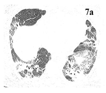
Figure 7a: Case 1 thorax CT image dated 2 April 2020. There are extensive patchy consolidated areas, more prominent in the lower zones.
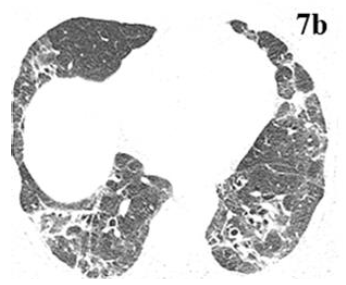
Figure 7b: Case 1 thorax CT image dated 20 April 2020. Significant regression was noted in the widespread consolidated areas observed in the lower zones compared with the previous CT.
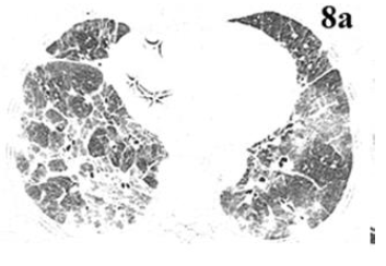
Figure 8a: Case 2 thorax CT image dated 9 April 2020. Widespread consolidated areas, reticular densities and fibrotic changes were noted in the lower zones of both lungs.
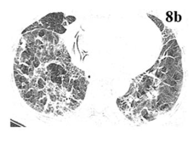
Figure 8b: Case2 thorax CT image dated 15 May 2020.Significant regression was observed in the parenchymal infiltration findings in both lungs. In addition, there is a significant decrease in fibrotic and atelectatic changes.
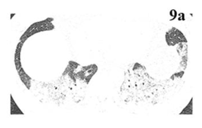
Figure 9a: Case 3 thorax CT image dated 18 April 2020. Secondary to diffuse infiltration in both lungs, a marked lack of ventilation was noted. Normal lung tissue cannot be observed in the lower zones.
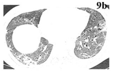
Figure 9b: Case thorax CT image dated 27 May 2020. Significant improvement in aeration is noted in both lung parenchymal areas. There is a significant decrease in the consolidation areas observed in the previous images.
3.2.3 Real Time Polymerase Chain reaction (RT-PCR) results: The RT-PCR was tested in the combined throat and nose swab specimens obtained from patients. The test dates and the results are illustrated in Figure 2, 4, and 6.
3.2.4 Adverse/Serious Adverse Events as Primary Safety indicators: Case 1 showed hypopotassaemia, hypernatremia, hyperglycaemia during stay in ICU. Hypopotassaemia was also seen in Case 2, with some hypertensive trend in vital and physical examination signs which were evaluated as part of the fluid balance signs and condition due to concomitant medical conditions, respectively. Conjunctival oedema and hyperaemia developed in the first day in ICU in Case 3, and it was resolved as the oxygenation is optimized. Agitation was the most prominent adverse event observed in this patient and it was accompanied by the incompliance with the treatments, these were seen on the 1st week of May. These were resolved gradually by the end of the combined treatment. Subcutaneous emphysema due to the patient-ventilator dyssynchrony was the finding of mid-May, lasting towards to end of May, and resolved with the improved general condition, hypopotassaemia and hypernatremia also resolved gradually. There were no combined treatment-related adverse or serious adverse events observed in any of the patients. Notably, there was no untoward adverse event or reaction related to the infusions before, during and after the MSC add-on treatment.

Figure 10a: Case 1- The counts of leucocytes and lymhocytes in hemogram assessment during the treatment. (Unit is K/uLfor both).

Figure 10b: Case 1- The thrombocytes counts in hemogram assessment during the treatment (Unit: K/uL).
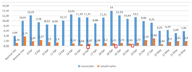
Figure 11a: Case 2- The counts of leucocytes and lymhocytes in hemogram assessment during the treatment.

Figure 11b: Case 2- The thrombocytes counts in hemogram assessment during the treatment.

Figure 12a: Case 3- The counts of leucocytes and lymhocytes in hemogram assessment during the treatment. (Unit is K/uLfor both).
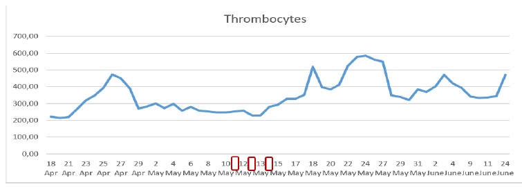
Figure 12b: Case 3- The thrombocytes counts in hemogram assessment during the treatment.
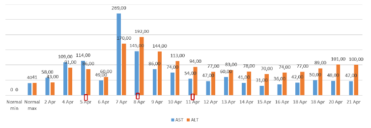
Figure 13a: Case 1- The liver function indicators AST, ALT levels (Unit: U/L for both).
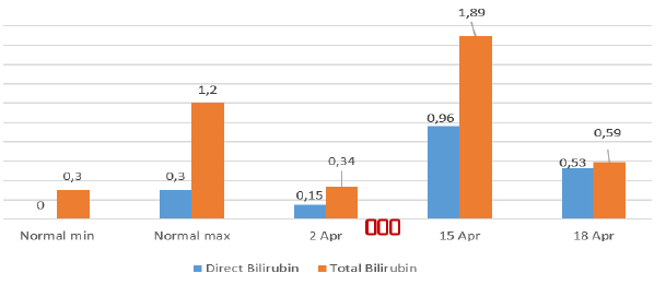
Figure 13b: Case 1- The liver function indicators total and direct bilirubin levels (Unit: mg/dL for both).
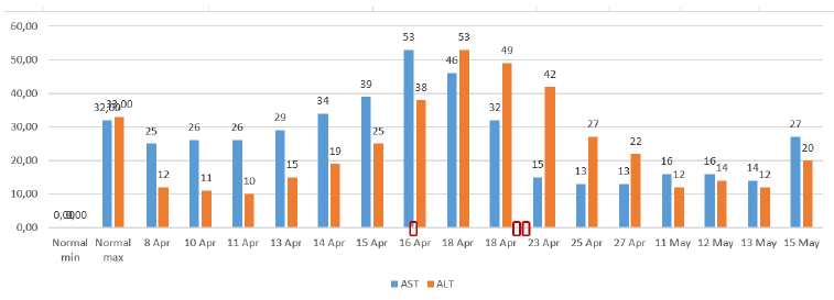
Figure 13c: Case 2- The liver function indicators AST, ALT levels.

Figure 13d: Case 2- The liver function indicators total and direct bilirubin levels.

Figure 13e: Case 3- The liver function indicators AST, ALT levels.

Figure 13f: Case 3- The liver function indicators total and direct bilirubin levels.
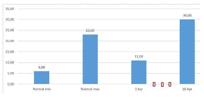
Figure 14a: Case 1- BUN levels as indicator of kidney function (Unit: mg/dL).

Figure 14b: Case 1- Blood creatinine levels as indicator of kidney function (Unit: mg/dL).
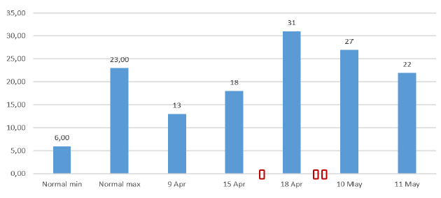
Figure 14c: Case 2- Blood Urea nitrogen levels as indicator of kidney function.
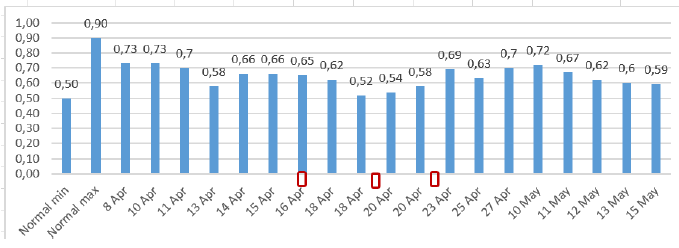
Figure 14d: Case 2- Blood creatinine levels as indicator of kidney function.

Figure 14e: Case 3- Blood Urea nitrogen (BUN) levels as indicator of kidney function.

Figure 14f: Case 3- Blood creatinine levels as indicator of kidney function.

Figure 15a: Case 1- Ferritin levels (ng/mL).
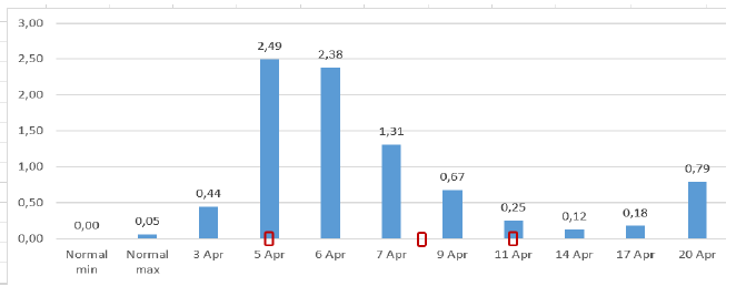
Figure 15b: Case 1- Procalcitonin levels (ng/mL).

Figure 15c: Case 1- Quantitative C-Reactive Protein (CRP) levels (mg/L).

Figure 15d: Case 2- Ferritin levels (ng/mL).
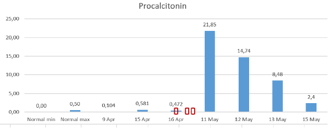
Figure 15e: Case 2- Procalcitonin levels.
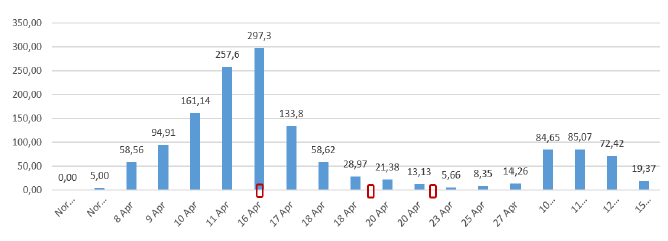
Figure 15f: Case 2- Quantitative C-Reactive Protein (CRP) levels.
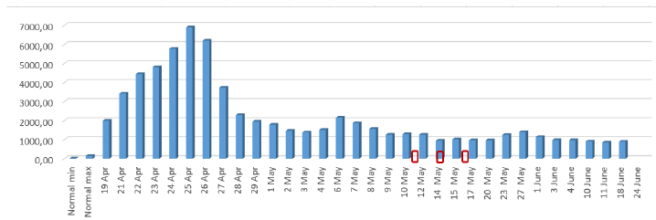
Figure 15g: Case 3- Ferritin levels.
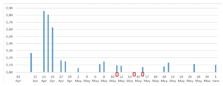
Figure 15h: Case 3- Procalcitonin levels.
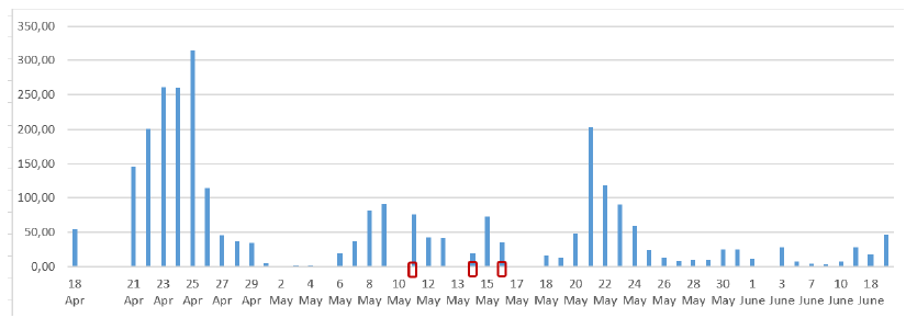
Figure 15i: Case 3- Quantitative C-Reactive Protein (CRP) levels.

Figure 16a: Case 1 - D-Dimer levels.
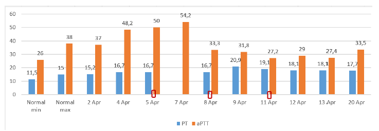
Figure 16b: Case 1 – PT and aPPT levels (seconds).

Figure 16c: Case 2 – D-Dimer levels.
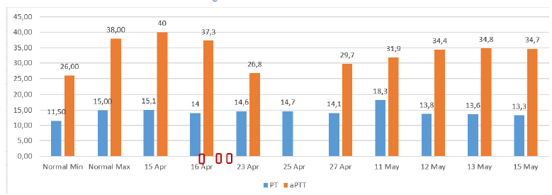
Figure 16d: Case 2 – PT and aPPT levels (seconds).

Figure 16e: Case 3 D Dimer levels.

Figure 16f: Case 3 – PT and aPTT levels.
3.2.5 Secondary efficacy indicators obtained from biological samples:
- White blood cells (WBC) or leucocytes, lymphocytes, red blood cells (RBC) and platelets levels in hemogram of the patients. These values are illustrated in Figures 10a-b, 11a-b, 12a-b. In all graphic figures, normal min and max colums are of normal value ranges. The red frame square refers to the MSC infusion days).
- Liver function tests of AST, ALT, direct and total bilirubin levels. These are illustrated in Figures 13 a-f.
- Kidney function tests of BUN, creatinine levels. These are illustrated in Figures 14a-f.
- Ferritin, Procalcitonin, CRP levels as inflammatory biomarkers are illustrated in Figures 15 a-i.
- Coagulation factors (D-Dimer, PT, aPTT) are illustrated in Figures 16a-i.
3.2.6 Blood, urine, tracheal aspirate culture results: The results of the specimen cultures were illustrated in Figure 2, 4 and 6 for Case 1, 2 and 3, respectively.
3. Discussion
So far, there is no specific treatment agent/s or protective vaccines are discovered, despite all the efforts. The countries affected by the novel pandemic have been recommending some treatment protocols consisted of already existing antiviral agents, immunomodulatory, anti-inflammatory agents with supportive treatments. Amongst the approaches, active vaccination by some vaccines [26] or passive vaccination by CP [27] are placed in the already existing treatment tools. Here, we explain the medical strategy our group followed, focusing on the combined treatment of critically ill COVID-19 patients with add-on MSCs therapy. COVID-19 shows complex pathogenesis and diversity of clinical outcomes. This is key to decide on the individual treatment regimes, particularly in severe and critically ill cases. Generally, in cases having severe respiratory distress with low oxygen saturation levels and findings of viral pneumonia images like multiple bilateral ground-glass opacities on chest CT scans, mechanical ventilation and ICU support may be needed. The advanced stage of the disease is characterised by cytokine storm and lymphopenia, and systemic clinical manifestations such as sepsis, septic shock, MOD, and ARDS [28, 29].
In our patients, we observed all signs in images confirming the criticality along with the concomitant chronic conditions, besides, each of them had their own progress confirming the varying clinical manifestations. For example, sepsis developed in the Case 1 and 3, but not in Case 2, MOD developed in Case 1, but not in Cases 2 and 3. MSC therapies have been under the scope to suppress cytokine storm and to prevent tissue damage and enhance alveolar tissue regeneration [30, 31]. In the cases we report combined treatment with add-on MSC therapy has been applied on a personalised manner depending on the individual conditions in means of days of infusions and the doses. Healing effects of the combined conventional treatment with add-on MSCs therapy was obvious in the improvement both in the oxygenation and thorax CT results, also in haematological parameters, coagulation capacity indicators, and in the decreased inflammatory markers of all cases. In parallel, the hepatic and renal functions were also improved in all cases. It is suggested that systematically infused MSCs could recover the pulmonary microenvironment, protect alveolar epithelial cells, intercept pulmonary fibrosis, and cure lung dysfunction and COVID-19 pneumonia [33].
Additionally, our safety data is an additional confirmatory evidence proving that there was no local or systemic adverse/serious adverse events or reactions developed. This was also an important data of a clinical trial [33]. As stated, we followed up three different cases showing varying clinical severity and treated them with add-on MSC therapy depending on the individual disease progress and treatment needs. That is why, we believe that MSC therapy contributed to the pulmonary improvement with immunomodulatory effect as we observed in Case 2, potential anti-inflammatory effect in Case 1 and antifibrotic effect as we suggested in Case 3.
We suggest that the MSC therapy is enhancing the conventional treatment outcome with its anti-fibrotic, immunomodulatory, anti-inflammatory and reparative effects modulating the healing in the impaired pulmonary microenvironment in parallel to the other published scientific data [34-38]. There is an urgent need to have the randomized controlled MSC treatment of COVID-19 studies published to get the golden evidence standard, nevertheless the value of the case reports on the relevant treatment option such as this one is incontrovertible, as it grounds in the individually tailored, personalized treatment approach.
5. Conclusion
We report three critically ill COVID-19 cases treated with add-on MSCs therapy during different courses of the disease progress in combination with the conventional therapies. We believe that this personalized therapeutic approach was resulted in cure and discharge of the patients. This paper also introduces promising safety data of the MSC therapy. We suggest that the MSC therapy could be a safe effective therapy method for novel coronavirus as add-on therapy when applied to the critically ill COVID-19 cases on a personalized manner.
References
- Yi Y, Lagniton P N P, Ye S, et al. COVID-19: what has been learned and to be learned about the novel coronavirus disease. Int. J. Biol. Sci 16 (2020): 1753-1766.
- Sun J, He W T, Wang L, et al. COVID-19: Epidemiology, Evolution, and Cross-Disciplinary Perspectives. Trends in Molecular Medicine 26 (2020).
- Xiea M, Chena O. Insight into 2019 novel coronavirus — An updated interim review and lessons from SARS-CoV and MERS-CoV. International Journal of Infectious Diseases 94 (2020): 119-124.
- Zhanga R, Wanga X, Nia L, et al. COVID-19: Melatonin as a potential adjuvant treatment. Life Sciences 250 (2020): 117583.
- Russell B, Moss C, George G, et al. Associations between immune-suppressive and stimulating drugs and novel COVID-19—a systematic review of current evidence. ecancer 14 (2020): 1022.
- Duan K, Liu B, Li C, et al. Effectiveness of convalescent plasma therapy in severe COVID-19 patients. Proc Natl Acad Sci S A 117 (2020): 9490-9496.
- Zhang C, Wu Z, Wen J, et al. Cytokine release syndrome in severe COVID-19: interleukin-6 receptor antagonist tocilizumab may be the key to reduce mortality. Int.J. Antimicrobial Agents 55 (2020): 105954.
- Shetty A K. Mesenchymal Stem Cell Infusion Shows Promise for Combating Coronavirus (COVID-19)- Induced Pneumonia. Aging and Disease 11 (2020): 462-464.
- Leng Z, Zhu Z, Hou W, et al. Transplantation of ACE2- Mesenchymal Stem Cells Improves the Outcome of Patients with COVID-19 Pneumonia. Aging and Disease 11 (2020): 216-228.
- Chen J, Hu C, Chen L, et al. Clinical Study of Mesenchymal Stem Cell Treatment for Acute Respiratory Distress Syndrome Induced by Epidemic Influenza A (H7N9) Infection: A Hint for COVID-19 Treatment. Engineering Engineering (Beijing) (2020).
- Wanga M, Luoc L, Bu H, et al. One case of coronavirus disease 2019 (COVID-19) in a patient co-infected by HIV with a low CD4+ T-cell count. Int J. Infectious Dis 96 (2020): 148-150.
- Singh S, Chakravarty T, Chen P, et al. Allogeneic cardiosphere-derived cells (CAP-1002) in critically ill COVID-19 patients: compassionate-use case series. Basic Research in Cardiology 115 (2020).
- Zheng G, Huang L, Tong H, et al. Treatment of acute respiratory distress syndrome with allogeneic adipose-derived mesenchymal stem cells: a randomized, placebo controlled pilot study. Respiratory research. 15 (2014): 39.
- Simonson O E, Mougiakakos D, Heldring N, et al. In Vivo Effects of Mesenchymal Stromal Cells in Two Patients with Severe Acute Respiratory Distress Syndrome. Stem Cells Translational Medicine 4 (2015): 1199-1213.
- Wilson J W, Liu K D, Zhuo H, et al. Mesenchymal stem (stromal) cells for treatment of ARDS: a phase 1 clinical trial. Lancet Respiratory Medicine 3 (2015): 24-32.
- Matthay M A, Calfee C S, Zhuo H, et al. Treatment with allogeneic mesenchymal stromal cells for moderate to severe acute respiratory distress syndrome (START study): a randomised phase 2a safety trial. Lancet Respiratory Medicine 7 (2019): 154-162.
- Atluri S, Manchikanti L, Hirsch J A. Expanded Umbilical Cord Mesenchymal Stem Cells (UC-MSCs) as a Therapeutic Strategy In Managing Critically Ill COVID-19 Patients: The Case for Compassionate Use Pain Physician 23 (2020): 71-83.
- Akkoc T. COVID-19 and Mesenchymal Stem Cell Treatment; Mystery or Not. the Advances in Experimental Medicine and Biology book series (2020): 1-10.
- Golchin A, Seyedjafari E, Ardeshirylajimi A. Mesenchymal Stem Cell Therapy for COVID-19: Present or Future. Stem Cell Rev Rep 16 (2020): 427-433.
- Yener D. "Türkiye'nin koronavirüsle mücadele politikasina 'Bilim Kurulu' yön veriyor". Ankara: Anadolu Ajansi (Anodolu Agent) (2020).
- TR ministry of health Covid-19 Information Page. Turkey Covid-19 Patient Table (2020).
- Regulation on Clinical Trials of Drugs and Biological Products. Official Gazette (2013).
- Official Circular on Stem Cell Studies (2018).
- Dai A, Baspinar O, Yesilyurt A, et al. Efficacy of stem cell therapy in ambulatory and nonambulatory children with Duchenne muscular dystrophy – Phase I–II.. Degenerative Neurological and Neuromuscular Disease 8 (2018): 63-77.
- Kabatas s¸ Civelek E, Inci C, et al. Wharton’s Jelly-Derived Mesenchymal Stem Cell Transplantation in a Patient with Hypoxic-Ischemic Encephalopathy: A Pilot Study Cell Transplantation 27 (2018): 1425-1433.
- Amanat F, Krammer F. SARS-CoV-2 vaccines: status report. Immunity 52 (2020): 583-589.
- Roback J D, Guarner J. Convalescent plasma to treat COVID-19: possibilities and challenges. JAMA (2020).
- Wu Z, McGoogan J M. Characteristics of and important lessons from the coronavirus disease 2019 (COVID-19) outbreak in China: summary of a report of 72314 cases from the Chinese Center for Disease Control and Prevention. JAMA 323 (2020): 1239-1242.
- Wang D, Hu B, Hu C, et al. Clinical characteristics of 138 hospitalized patients with 2019 novel coronavirus-infected pneumonia in Wuhan, China. JAMA 323 (2020): 1061-1069.
- Rogers C J, Harman R J, Bunnell B A, et al. Minev, Rationale for the clinical use of adipose-derived mesenchymal stem cells for COVID-19 patients. JAMA 323 (2020): 1061-1069.
- Broxmeyer H E, Parke G C. Impact of COVID-19 and Future Emerging Viruses on Hematopoietic Cell Transplantation and Other Cellular Therapies. Stem Cells and Development 29 (2020): 10.
- Bari E, Ferrarotti I, Saracino L, et al. Mesenchymal Stromal Cell Secretome for Severe COVID-19Infections: Premises fort he Therapeutic Use. Cells 9 (2020): 924.
- Leng Z, Zhu R, Hou W, et al. Transplantation of ACE2- Mesenchymal stem cells improves the outcome of patients with COVID-19 pneumonia. Aging and Disease 11 (2020): 216.
- Wu C, Chen X, Cai Y, et al. Risk factors associated with acute respiratory distress syndrome and death in patients with coronavirus disease 2019 pneumonia in Wuhan, China. JAMA Intern. Med 180 (2020): 1-11.
- Zhou B, She J, Wang Y, et al. Utility of ferritin, procalcitonin, and C-reactive protein in severe patients with 2019 novel coronavirus disease. Preprint from Research Square (2020).
- Luo X, Yan W, Zhou X, et al. Prognostic value of C-reactive protein in patients with COVID-19. Clinical Infectious Diseases (2020).
- Liu T, Zhang J, Yang Y, et al. The potential role of IL-6 in monitoring severe case of coronavirus disease 2019. medRxiv (2020).
- Zhou F, Yu T, Du R, et al. Clinical course and risk factors for mortality of adult inpatients with COVID-19 in Wuhan, China: a retrospective cohort study. Lancet 395 (2020): 1054-1062.


 Impact Factor: * 5.3
 Acceptance Rate: 75.63%
  Time to first decision: 10.4 days
  Time from article received to acceptance: 2-3 weeks
