Bowel Lymphoma Causing Multiple Intussusceptions In Adult- Case Report and Review of Literature
Gita Devi, MD*, Lokesh Singh, MD, Uma Debi, MD, M S Sandhu, MD
Radiodiagnosis and Imaging, PGIMER Chandigarh, India
*Corresponding Author: Dr. Gita Devi, MD, Radiodiagnosis and Imaging, PGIMER Chandigarh, India
Received: 28 May 2020; Accepted: 19 June 2020; Published: 15 July 2020
Article Information
Citation: Gita Devi, Lokesh Singh, Uma Debi, Sandhu MS. Bowel Lymphoma Causing Multiple Intussusceptions In Adult- Case Report and Review of Literature. Archives of Clinical and Medical Case Reports 4 (2020): 707-711.
View / Download Pdf Share at FacebookAbstract
Intussusception in adults is rarer than children and treatment in most cases in adults is surgical. Multiple Intussusceptions in our case with subacute intestinal obstruction and presence of abdominal lymphadenopathy with diffuse bowel wall thickening warrants non-surgical treatment. Presence of multiple intussusceptions is rare findings. We present a case of female aged 35 years presented with palpable abdominal lump associated with loss of weight and appetite. CECT abdomen showed abdominal lymphadenopathy and multisegmental small bowel thickening maximum upto 1.5 cm. Some segments of bowel also show aneurysmal dilatation associated with asymmetrical mural thickening. Telescoping of proximal bowel segment into adjacent distal bowel is seen in at least two places in ascending colon region (colocolic intussusceptions) and another in ileal loops (ileo-ileal). FNAC from the lymph nodes revealed non hodgkins lymphoma.
Keywords
<p>Adult intussusceptions; Asymmetrical bowel wall thickening; Non Hodgkin’s lymphoma</p>
Article Details
1. Introduction
Intussusception was first defined in 1674 as telescoping of a bowel loop with its mesenteric fold (intussusceptum) into the lumen of contiguous part of bowel (intussuscipien) [1]. Intussusception is much rare in adults as compared to children and accounts for only 5% of cases. Detectable underlying cause of intussusceptions however is more common in adults and causes range from idiopathic to benign and malignant aetiologies [1]. Most common site for adult intussusceptions is at junctions of fixed and freely moving segments of bowel [1].
2. Case Report
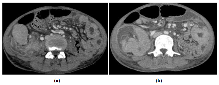
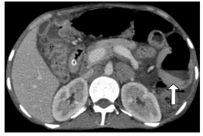
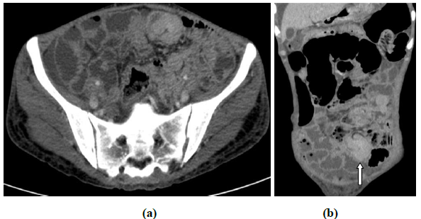
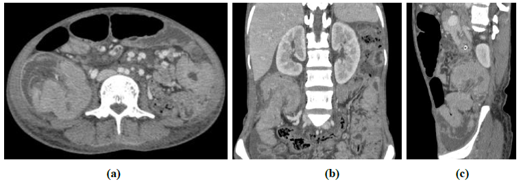
Female aged 35 years presented with palpable abdominal lump associated with loss of weight and appetite. CECT abdomen was done which showed multisegmental small bowel thickening maximum measuring up to 1.5 cm (Figure 1). Some segments of bowel also showed aneurysmal dilatation associated with asymmetrical mural thickening (Figure 2). Telescoping of proximal bowel segment into adjacent distal bowel was seen in at least two places in ascending colon region (colocolic intussusceptions) and another in ileum(ilioileal) loops (Figure 3 and 4). These were long segmental bowel wall thickenings. Mild dilatation of proximal bowel loops consistent with subacute intestinal obstruction was present. Enlarged lymph nodes noted in perigastric region, mesentery and in retroperitoneum. Lymph nodal mass observed in the perigastric location infilterating full thickness of stomach wall. Multiple of these lymph nodes showed necrosis [5]. FNAC from the lymph nodes revealed non hodgkins lymphoma (Figure 5).

Figure 1: Axial CT images showing multisegmental bowel wall thickening involving small and large bowel loops (a and b).

Figure 2: Axial CECT image showing aneurismal dilatation of jejunum (left side arrow) associated with asymmetrical mural thickening. Proximal and distal to this loop is not dilated.

Figure 3: CECT Axial (a) and coronal (b) images showing ileioileal intussusception (white arrows) associated with asymmetrical mural thickening.

Figure 4: CECT Axial (a), coronal (b) and sagittal(c) images showing long segmental colocolic intussusception associated with asymmetrical mural thickening.

Figure 5: CECT axial (a, b) and coronal (c) images showing lymph nodes in perigastric region, mesentery and in retroperitoneum. Lymph nodal mass in perigastric location infilterating the full thickness of stomach wall (a). Multiple of lymph nodes with necrosis (b and c).
3. Discussion
According to the site of origin intussusception have been classified in four types namely enteric, ileocolic, ileocaecal and colonic [2]. Intussusception is also classified based on aetiology as benign, malignant or idiopathic. Marinis et al. [3] found a case of ileo-colic intussusception due to small B-cell (Burkitt-like) NHL of ileum in 29 year male. Intussusceptions in colon are more likely malignant [3]. Symptoms of intussusceptions in adult vary and are non-specific. Periodic and intermittent pain is common symptom patients present with. CT scan is the investigation of choice for making definite diagnosis in patients with non-specific pain. In adults laprotomy is the treatment of choice as there is underlying cause in maximum of the cases and especially in when it presents as acute abdomen.
Resection is advised without attempting reduction when the bowel is inflamed, ischemic or in colo-colic intussusceptions [4].
Surgical resection without reduction should be preferred treatment in adult intussusception and only reduction of intussusceptions should be done in post –traumatic and idiopathic cases [2]. Yalamarthi S and Smith RC in their Studies of four cases of intussusceptions found benign tubulovillous adenoma of jejunum, lipoma of duodenum, large Caecal villous adenoma and ulcerated Meckel’s diverticulum as the causes of adult intussusceptions [2]. Hameed T et al. reported a case of acute ileo-colic intussusceptions in a middle aged patient due to inflammatory myofibroblastic tumor of ileum, a rare tumor in adults [5] and Shephered T et al. reported colocolic intussusceptions due to lipoma as the cause of leading point [6]. These small lesions present as submucosal masses. These masses are pulled ahead by peristalsis due to which affected segment of bowel is prolapsed into the adjacent usually normal segment. Rarely lymphangiectasia of intestinal wall can also present with intussusception [7].
In our case NHL is the cause of intussusceptions involving right colon and small bowel where the leading point is asymmetrical mural thickening associated with abdominal lymphadenopathy. Adult intussusceptions accounts for approximately 5% of all cases of and 1-5% of intestinalobstruction. Adult intussusceptions can be idiopathic (8-20%) or secondary in which underlying cause is organic lesions. These are inflammatory bowel disease, Meckel’s diverticulum, benign and malignant lesions, metastatic neoplasms and iatrogenic causes like due to intestinal tubes or post-surgical adhesions. Reduction is safe in case of benign aetiologies and is preferred prior to surgery to limit the extent of resection and avoid short bowel syndrome [3].
4. Conclusion
Adult intusussceptions are rare and present with nonspecific symptoms. Computed tomography plays an important role in diagnosis, treatment planning and follow up of these cases.
References
- Brigg JH, Mckean D, Palmer JS, et al. Transient small bowel intussusceptions in adults: as overlooked feature of coelic disease. BMJ Case Rep (2014): bcr2013203156.
- Yalamarthi S, Smith RC. Adult intussusception: care reports and review of literature. Postgrad Med J 81 (2005): 174-177.
- Marinis A, Yiallourov A, Samanides L, et al. Intussusception of the bowel in adults: A review. World J Gastroenterol 15 (2009): 407-411.
- Begos DG, Sandor A, Moldin IM. The diagnosis and management of adult intussusception. Am j Surg 121 (1971): 531-535.
- Hameed T, Singh M, Nizam A, et al. Acute Intestinal Obstruction Due to Ileocolic Intussusception in an Adult; A Rare Presentation of Inflammatory Myofibroblastic Tumor. Am J Case Rep 21 (2020): 1-5.
- Shepherd T, Wazir M, Cover J. Rare case of adult colocolic intussusceptions. BMJ Case Rep 13 (2020): e232761.
- Zafar Y, Gondi KT, Tamimi T, et al. Primary Intestinal Lymphangiectasia Causing Intussusception and Small Bowel Obstruction. ACG Case Rep J 6 (2019): e00233.


 Impact Factor: * 5.3
 Acceptance Rate: 75.63%
  Time to first decision: 10.4 days
  Time from article received to acceptance: 2-3 weeks
