Case Report: Histological Evaluation of Deep Tissue Remodeling After Extended Mode of Fractional RF Microneedling
Chien-Ming Chen MD1* and Jieyang Jhuang MD2
1Belleesse Dermatological Aesthetic Clinic, Zhongshan District, Taipei City, Taiwan
2Department of Pathology, Mackay Memorial Hospital, Zhongshan North Road, Zhongshan District, Taipei City, Taiwan
*Corresponding Author: Chien-Ming Chen, Belleesse Dermatological Aesthetic Clinic, Zhongshan District, Taipei City, Taiwan.
Received: 11 August 2025; Accepted: 19 August 2025; Published: 29 August 2024
Article Information
Citation: Chien-Ming Chen, Jieyang Jhuang. Case Report: Histological Evaluation of Deep Tissue Remodeling After Extended Mode of Fractional RF Microneedling. Archives of Clinical and Medical Case Reports. 9 (2025): 175-180.
View / Download Pdf Share at FacebookAbstract
Fractional radiofrequency (RF) microneedling is commonly used for skin and body rejuvenation by inducing controlled thermal micro-injuries that stimulate collagen and elastin production. However, conventional techniques often cause discomfort, especially when targeting deeper tissues. This case report evaluates the histological effects of the Exion Fractional RF device using its Extended Mode, which creates a thermal gradient reaching deeper layers without full needle penetration. A 40-yearold male subject (BMI 22.9) received a single-pass treatment to the face and abdomen, with insulated microneedles delivering monopolar RF energy at varying depths (1.0–4.0 mm) and intensities (60% and 100%). Biopsies were taken immediately post-treatment, and on Days 7 and 30. Histological analysis using H&E and Masson's trichrome staining revealed a consistent 4-mm thermal gradient in abdominal tissue, with evidence of collagen coagulation and remodeling extending beyond needle depth, while preserving the integrity of the epidermis. By Day 30, early neocollagenesis and signs of adipocyte apoptosis, including membrane disruption and fibrotic changes, were noted. These findings suggest that the Extended Mode enables effective deep tissue remodeling and adipose targeting while minimizing epidermal disruption.
Keywords
<p>Fractional Radiofrequency; Microneedling; Skin rejuvenation; Histology; Conventional techniques; Deep tissue remodeling</p>
Article Details
1. Introduction
Microneedling paired with radiofrequency (RF) effectively rejuvenates the face and body [1]. This innovative procedure delivers monopolar RF energy through needles that penetrate the skin at specific depths, stimulating collagen and elastin production [2]. While highly effective, the treatment's mechanism involves creating controlled micro-injuries through needle penetration, which can cause pain to varying extents.
Exion Fractional RF technology introduces a solution to address patient discomfort during microneedling treatments with its innovative Extended Mode feature. This technology combines monopolar RF with AI-controlled energy delivery while leveraging the Extended Mode to generate a thermal gradient that reaches up to 8 mm in depth, extending 4 mm beyond the maximum needle penetration depth of 4 mm [3,4]. By effectively targeting subdermal layers without physically piercing those depths, the Extended Mode minimizes patient discomfort while achieving optimal results.
The gradient is created by monopolar RF energy, with a neutral electrode positioned on the patient's back. When the Extended Mode is engaged, the RF pulse is delivered to the needle tip, and the energy flows toward the neutral electrode, creating a consistent 4mm thermal gradient beneath the needle [5]. In contrast, without Extended Mode, RF energy is confined to the tissue directly surrounding the needle tip, limiting the effective depth of thermal stimulation to the depth of mechanical insertion.
This case report observes the histological changes induced by the device's Extended Mode at depths beyond the physical needle insertion, while also demonstrating its mechanism of action.
2. Methods
This case report describes histological observations from a single subject (n=1, male, 40 years old, BMI 22.9) who underwent a single-pass fractional RF treatment using the EXION device (BTL Industries Inc., Boston, MA). This case was managed by the ethical principles outlined in the 1975 Declaration of Helsinki. The treatment was applied to the facial and abdominal areas.
The subject was required to complete two (2) follow-up visits scheduled for 7 days and 30 days post- treatment. Adverse events were closely monitored throughout the case study duration.
The preauricular and abdominal areas were cleansed before therapy initiation, and digital photographs with marked biopsy sites were taken. Tissue samples were obtained using a 3 mm punch biopsy immediately after the treatment, 7 days post-treatment, and 30 days post-treatment. The samples were sectioned and stained to facilitate visualization and microscopic examination. The specimens were stained with hematoxylin-eosin (H&E) and Masson's trichrome (MT) following standard staining protocols.
In the facial area, the treatment was conducted at 60% RF intensity, with a needle penetration depth of 1.0 mm, and with Extended Mode disabled. The sample for histological evaluation was taken immediately following the treatment. In the abdominal area, the treatment was performed with Extended Mode enabled, using two RF intensity levels (60% and 100%) and four needle penetration depths (1.0 mm, 1.5 mm, 2.5 mm, and 4.0 mm). Samples for histological evaluation were taken immediately following the treatment, 7 days post-treatment, and 30 days post- treatment.
3. Results
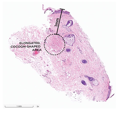
Histological samples stained with H&E showed specific morphological changes in the treated facial area. A distinctive, elongated, and cocoon-shaped area was observed, indicating the coagulation of tissue proteins, such as collagen (Figure 1). These localized changes were consistent with standard RF microneedling (with Extended Mode disabled), as no evidence of a broader thermal gradient or deeper tissue remodeling was observed.
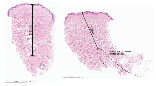
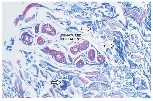
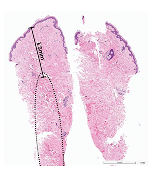
Histological images of the abdominal area, stained with H&E and MT, demonstrated a 4-mm thermal gradient characterized by a localized zone of collagen denaturation and dermal coagulation. The epidermis remained unaffected by the RF energy due to the insulated needles, confining the effect to the targeted tissue depth (Figures 2-4). Disorganized, fragmented, and swollen collagen fibers characterized the collagen denaturation zone. On Day 0, there was no evidence of collagen remodeling. However, clear signs of acute inflammation, indicative of the initial phase of the healing cascade, were visible. This was marked by the presence of interstitial spaces between the collagen fibers, representing tissue edema. By Day 30, the tissue transitioned into the proliferative phase of healing, with early signs of collagen remodeling becoming more prominent, marked by a denser and better-organized matrix. See Figure 2. Regardless of the needle penetration depth, the thermal gradient consistently reached a depth of 4 mm, as evidenced by the changes in collagen organization within the deeper dermal layers. While the histological sections were prepared to a maximum depth of 4 mm, the thermal gradient was consistently visible across the full extent of the analyzed tissue, indicating its effect throughout the examined depth beneath the needle tip.
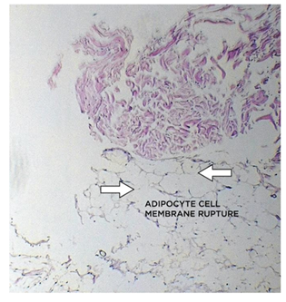
In addition, the images showed morphological changes in adipose tissue, suggesting a potential effect on fat reduction. At Day 0, adipocytes showed early signs of membrane disruption due to RF energy exposure, as illustrated in Figure 5. By Day 7, there was clear evidence of ruptured adipocytes, which appeared as irregularly shaped or fragmented cells, indicating a loss of structural integrity. This was accompanied by a marked loss of adipocyte architecture, with the typical clear, rounded fat cells replaced by disorganized tissue, along with fibrotic changes in the fat layer beneath the eccrine coil.
Fibrotic remodeling, characterized by the presence of dense connective tissue primarily composed of collagen, was observed in areas where adipose tissue structure appeared disrupted. The typical vacuolated morphology of adipocytes was absent, and collagen fibers were identifiable through dense eosinophilic staining.
No treatment-related adverse events or side effects were observed, and the punch biopsy sites healed normally.
4. Discussion
This case report describes histological changes observed following microneedling RF treatment using the device's Extended Mode with insulated microneedle tips. Histological evaluation revealed morphological changes extending up to 4 mm beyond the set penetration depth of the microneedle tip, suggesting that the Extended Mode facilitates tissue effects that extend beyond the physical reach of the needle itself.
Previous studies have shown that microneedle penetration combined with RF energy delivery activates the body's natural healing response, thereby initiating neocollagenesis [6]. These studies included histological assessments of thermally treated tissue, which demonstrated clear evidence of collagen coagulation and denaturation, followed by complete replacement with new collagen fibers observed 90 days post-treatment [7]. RF-induced heat leads to the denaturation of existing collagen [8], while the resulting mechanical and thermal stimuli activate the three phases of neocollagenesis: inflammation, proliferation, and remodeling [9,10]. In our case, the observed depth of collagen remodeling suggests that the Extended Mode is able to deliver energy to the deeper dermal layers. Furthermore, the remodeling phase, which involves the maturation of new collagen, can persist for several months to over a year post-treatment [11]. Since collagen remodeling is a gradual process, the full extent of collagen increase cannot be accurately assessed at Day 30. Therefore, our focus on early structural changes provides a foundation for understanding the long- term benefits of Extended Mode treatment. Moreover, we hypothesize that if different staining methods had been used to evaluate the samples, we might have observed immature type III collagen, an early sign of neocollagenesis.
The use of Extended Mode allowed for deeper delivery of RF energy, effectively targeting the dermis and subcutaneous fat layers while maintaining the integrity of the epidermis. This precise targeting minimizes surface damage, reducing downtime and the risk of complications while promoting collagen remodeling and tissue regeneration in the deeper layers [12].
Previous studies have suggested that RF energy induces adipocyte apoptosis [13-15]. Although direct apoptotic markers were not assessed in this case report, the morphological features were consistent with adipocyte disruption, particularly in the subcutaneous fat tissue, where early thermal effects were observed. By Day 0, signs of thermal injury were evident in the subcutaneous fat layer, with adipocytes showing structural changes indicative of early damage. However, no significant fibrosis was present at this stage. In follow-up evaluations, clear signs of adipocyte breakdown and fibrotic tissue replacement were observed. This transition may reflect the initiation of a long-term fat reduction process, in which damaged adipocytes are gradually replaced by fibrotic tissue, contributing to improved structural integrity and skin firmness [16].
Fibrosis refers to the formation of excess connective tissue, primarily collagen, as part of the body’s healing response to injury [17]. Depending on the context, fibrosis can be either beneficial or harmful. When induced in a controlled manner, such as with microneedling RF, fibrosis can be advantageous. In this case, fibrosis contributes to tissue remodeling and structural improvement, particularly in the dermis and subcutaneous fat. The controlled fibrosis seen with RF treatment promotes neocollagenesis and structural adipocyte disruption, aiding in skin tightening and body contouring [18,19].
A key strength of this case report is the histological analysis of human tissue, with multiple samples varying in needle penetration depth, RF intensity, and sampling time points from a single subject. However, this report also contains several limitations. First, the follow-up period was restricted to 30 days post-treatment, which may not represent the long-term remodeling of collagen. Another limitation is the depth of sample collection, as it did not extend far enough to capture the full thermal gradient beyond 4 mm. Furthermore, only insulated microneedle tips were used, highlighting the need for future studies to incorporate both insulated and non-insulated tips to provide a broader understanding of the procedure’s impact on various tissue depths and thermal distributions. Future investigations should also aim to include a larger sample size, longer follow- up periods to monitor long-term collagen formation, deeper sample collection, and varied needle types to provide a more comprehensive understanding of the treatment’s efficacy and mechanism of action.
5. Conclusions
Histological analysis confirmed the effectiveness of Exion Fractional RF treatment with the Extended Mode in promoting deep tissue remodeling. The findings indicate that the Extended Mode consistently produces a 4-mm thermal gradient, regardless of needle penetration depth. Immediate thermal effects, such as collagen coagulation, were evident in tissue samples. Over the course of the treatment, early signs of active tissue remodeling were observed, including neocollagenesis and evidence of structural adipocyte disruption in the subcutaneous fat. These histological changes were accompanied by reduced inflammation and improved structural integrity of the dermis. The observed adipose remodeling suggests the initiation of a long-term tissue contouring process.
6. Ethical Approval
The authors confirm that the ethical policies of the journal, as noted on the journal’s author guidelines page have been adhered to. The subject voluntarily participated and signed a written informed consent.
References
- Syder NC, Chen A, Elbuluk N. Radiofrequency and Radiofrequency Microneedling in Skin of Color: A Review of Usage, Safety, and Efficacy. Dermatol Surg 49 (2023): 489.
- Weiner SF. Radiofrequency Microneedling: Overview of Technology, Advantages, Differences in Devices, Studies, and Indications. Facial Plast Surg Clin N Am 27 (2019): 291-303.
- Gupta J, Park SS, Bondy B, et al. Infusion pressure and pain during microneedle injection into skin of human subjects. Biomaterials 32 (2011): 6823-6831.
- Tanaka Y. Nonsurgical facial tightening after a fractional noninsulated microneedling radiofrequency treatment in Asians. Dermatol Rev 1 (2020): 20-26.
- Shellock FG. Radiofrequency energy induced heating of bovine capsular tissue: in vitro assessment of newly developed, temperature-controlled monopolar and bipolar radiofrequency electrodes. Knee Surg Sports Traumatol Arthrosc 10 (2002): 254-259.
- Meyer PF, de Oliveira P, Silva FKBA, et al. Radiofrequency treatment induces fibroblast growth factor 2 expression and subsequently promotes neocollagenesis and neoangiogenesis in the skin tissue. Lasers Med Sci 32 (2017): 1727-1736.
- Wang H, Hamblin MR, Zhang Y, et al. Histological evaluation of monopolar and bipolar radiofrequency microneedling treatment in a porcine model. Lasers Surg Med 56 (2024): 288-297.
- Manuskiatti W, Pattanaprichakul P, Inthasotti S, et al. Thermal Response of In Vivo Human Skin to Fractional Radiofrequency Microneedle Device. BioMed Res Int (2016): 6939018.
- Schmitt L, Marquardt Y, Amann P, et al. Comprehensive molecular characterization of microneedling therapy in a human three-dimensional skin model. PLOS ONE 13 (2018): e0204318.
- Liebl H, Kloth LC. Skin cell proliferation stimulated by microneedles. J Am Coll Clin Wound Spec 4 (2012): 2-6.
- Tan MG, Jo CE, Chapas A, et al. Radiofrequency Microneedling: A Comprehensive and Critical Review. Dermatol Surg 47 (2021): 755.
- Bernardy J. Investigation of Histological Changes Induces by a Novel Fractional Radiofrequency Device for Skin Rejuvenation in Porcine Skin Tissue (2023).
- Vale AL, Pereira AS, Morais A, et al. Effects of radiofrequency on adipose tissue: A systematic review with meta-analysis. J Cosmet Dermatol 17 (2018): 703-711.
- McDaniel D, Fritz K, Machovcova A, et al. A focused monopolar radiofrequency causes apoptosis: a porcine model. J Drugs Dermatol JDD 13 (2014): 1336-1340.
- Boisnic S, Divaris M, Nelson AA, et al. A clinical and biological evaluation of a novel, noninvasive radiofrequency device for the long-term reduction of adipose tissue. Lasers Surg Med 46 (2014): 94-103.
- Kruglikov IL, Scherer PE. Skin aging: are adipocytes the next target? Aging 8 (2016): 1457-1469.
- Jun JI, Lau LF. Resolution of organ fibrosis. J Clin Invest 128 (2018): 97-107.
- Kleaveland KR, Moore BB, Kim KK. Paracrine functions of fibrocytes to promote lung fibrosis. Expert Rev Respir Med 8 (2014): 163-172.
- Baek G, Kim MH, Jue MS. Efficacy of microneedle radiofrequency therapy in the treatment of senile purpura: A prospective study. Skin Res Technol 28 (2022): 856-864.


 Impact Factor: * 5.3
 Acceptance Rate: 75.63%
  Time to first decision: 10.4 days
  Time from article received to acceptance: 2-3 weeks
